Nodule detection

a computed tomography and nodule technology, applied in the field of nodule detection, can solve the problems of difficult detection of nodules, incorrect identification of characteristics of nodules, and difficulty in nodule detection

Inactive Publication Date: 2005-11-24
SAMSUNG ELECTRONICS CO LTD
View PDF8 Cites 11 Cited by
  • Summary
  • Abstract
  • Description
  • Claims
  • Application Information

AI Technical Summary

Benefits of technology

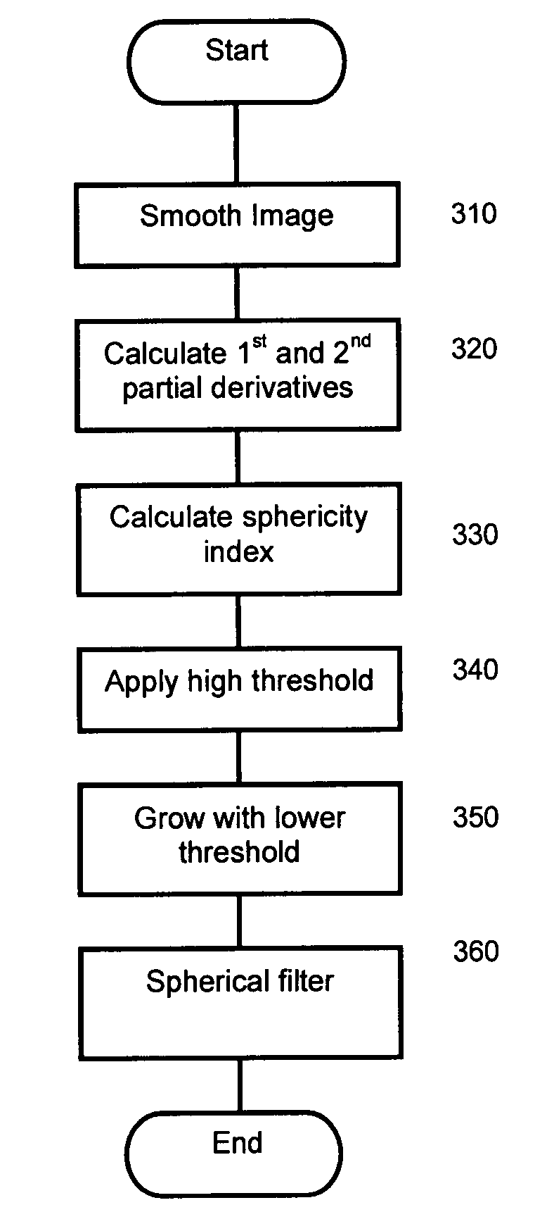
[0009] According to an embodiment of the present invention, there is provided a method of detecting a nodule in a three-dimensional scan image, comprising calculating a sphericity index for each point in the scan image relative to surrounding points of similar intensity, applying a high sphericity threshold to the sphericity index to obtain a candidate nodule region, and then performing region-growing from the candidate region using a relaxed sphericity threshold to identify an extended region including less spherical parts connected to the candidate region. The extended region may be provided for display and / or for subsequent processing to calculate physical characteristics and / or to perform automatic diagnosis. In an embodiment, diagnosis may be performed by a radiologist on the basis of the enhanced image.

Problems solved by technology

Nodule detection is one of the more challenging tasks in medical imaging.
Nodules may be difficult to detect on CT scans because of low contrast, small size, or location of the nodule within an area of complicated anatomy.
A spherical model may be useful to segment nodules from surrounding objects, but if the result is to incorrectly identify the nodule as a sphere and to discard non-spherical portions of the nodule, the characteristics of the nodule may also be incorrectly identified.

Method used


Image

  • Nodule detection
  • Nodule detection
  • Nodule detection

Examples


Embodiment Construction

[0024] CT Image

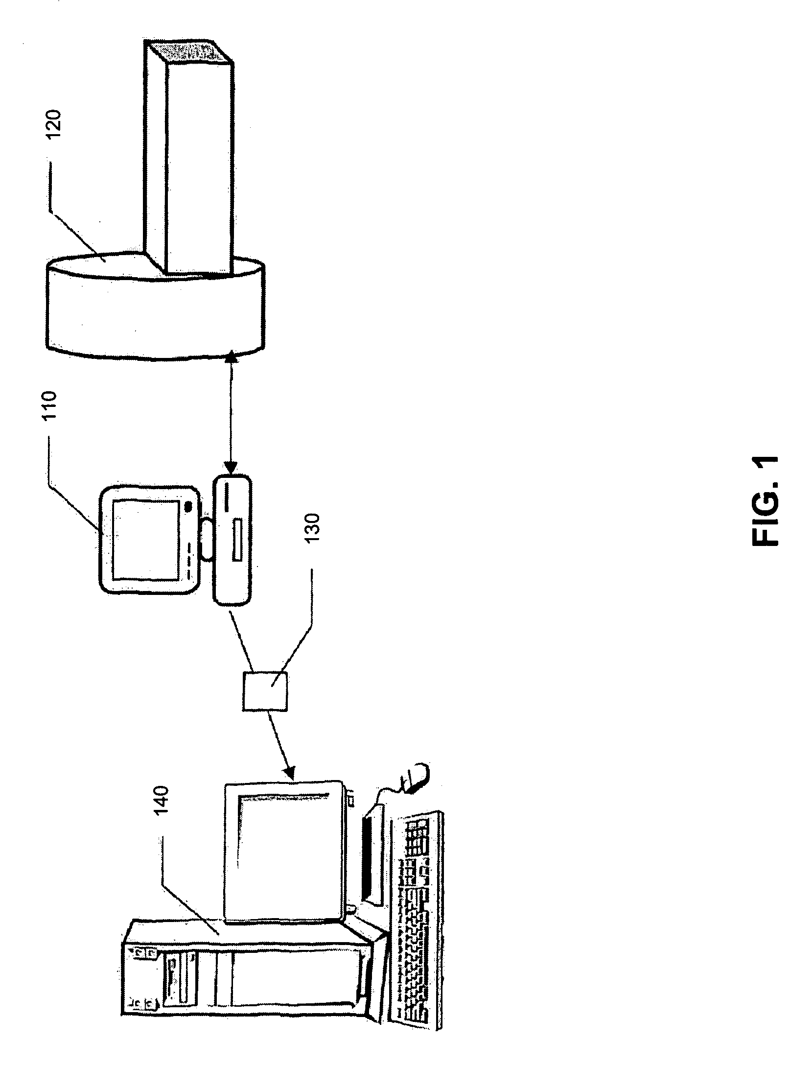
[0025] Each embodiment is performed on series of CT image slices obtained from a CT scan of the chest area of a human or animal patient. Each slice is a 2-dimensional digital grey-scale image of the x-ray absorption of the scanned area. The properties of the slice depend on the CT scanner used; for example, a high-resolution multi-slice CT scanner may produce images with a resolution of 0.5-0.6 mm / pixel in the x and y directions (i.e. in the plane of the slice). Each pixel may have 32-bit grayscale resolution. The intensity value of each pixel is normally expressed in Hounsfield units (HU). Sequential slices may be separated by a constant distance along the z direction (i.e. the scan separation axis); for example, by a distance of between 0.75-2.5 mm. Hence, the scan image is a three-dimensional (3D) grey scale image, with an overall size depending on the area and number of slices scanned.

[0026] The present invention is not restricted to any specific scanning techni...


PUM


Abstract

A method of detecting a nodule in a three-dimensional scan image comprises calculating a three-dimensional sphericity index for each point in the scan image (310-330), applying a high sphericity threshold to the sphericity index (340) to obtain a candidate nodule region, and then performing region-growing (350) from the candidate region using a relaxed sphericity threshold to determine an extended region including less spherical parts connected to the candidate region. Optionally, spherical filtering may be applied to the image by matching the spherical filter to the extended region.

Description

RELATED APPLICATIONS [0001] This application claims the benefit of the filing date of GB Patent Application No. 0411284.3, filed May 20, 2004, which is incorporated herein by reference in its entirety. FIELD OF THE INVENTION [0002] The present invention relates to a method of detecting nodules in computed tomography (CT) images, and particularly but not exclusively for detecting nodules in CT images of the lung. The method may be implemented using a computer, and the invention encompasses software and apparatus for carrying out the method. BACKGROUND OF THE INVENTION [0003] The mortality rate for lung cancer is higher than that for other kinds of cancers around the world. Detection of suspicious lesions in the early stages of cancer can be considered the most effective way to improve survival. Nodule detection is one of the more challenging tasks in medical imaging. Nodules may be difficult to detect on CT scans because of low contrast, small size, or location of the nodule within a...

Claims


Application Information

Patent Timeline
Patent Type & Authority Applications(United States)
IPC IPC(8): A61B6/03G06T7/00G06T7/60G06V10/46
CPCA61B6/032G06K9/48G06K2209/05G06T2207/30064G06T7/604G06T2207/10081G06T2207/20141G06T7/0012G06T7/64G06T7/187G06V10/46G06V2201/03G06T5/10A61B6/03G06V10/10
Inventor DEHMESHKI, JAMSHID
Owner SAMSUNG ELECTRONICS CO LTD
Why Eureka
Try Eureka