Atlas reporting

a radiographic examination and atlas technology, applied in the field of diagnostic radiographic examinations, can solve the problems of dragging, dragging, and dragging, and achieve the effect of improving the policy decision making process for research funding and healthcare money allocation, and rapid and accurate billing

Inactive Publication Date: 2006-12-07
ATLAS REPORTING
View PDF9 Cites 80 Cited by
  • Summary
  • Abstract
  • Description
  • Claims
  • Application Information

AI Technical Summary

Benefits of technology

[0015] In an embodiment of the invention storage of disease codes with the anatomic drawings allows automated retrieval and documentation of disease prevalence, morbidity, and response to treatment. Creating a similar database from “free text reporting” is difficult, if not impossible. This embodiment could be used to automatically gather information comparing the effectiveness of different treatments of disease. It may provide important epidemiological data to observing authorities such as the Centers for Disease Control. Compiling data from multiple healthcare centers using the invention could improve policy decisions for research funding and healthcare money allocations. Where a surprising or unusual occurrence of a condition is observed, the immediate coding by the embedded means of an embodiment of the invention serves to alert the authorities of the condition. Optionally, the alerts may be automated such that the statistical occurrence across a population may be charted, alerting the authority to an outbreak of a disease in numbers greater than would be expected across the monitored population.
[0016] A further embodiment of the invention allows correlation of the billable work (Current Procedural Terminology code or CPT code) involved in the medical imaging procedure with the clinical presentation (provider's reasons for having ordered the examination) of the patient and eventual ICD-10 code. Reimbursement for medical image examinations from Medicare and third party payers is dependant on appropriate correlation of the examination with the patient's clinical presentation. The reimbursement level may also differ depending on the diagnosis that caused the clinical problems. In this embodiment of the invention, embedded code allows correlation of the CPT code with the ICD-10 to effect rapid and accurate billing. Optionally, the embedded coding may interact with a database to inform the radiologist of precedent threshold questions being raised by the payer. In use of such an embodiment, the radiologist's suggested follow-up regimen may be tailored to reflect the payer's requirements.

Problems solved by technology

Verbal dictation, on the other hand, a left-brain language function, is kept to a minimum, as it is tedious, linear, and tiring.

Method used


Image

  • Atlas reporting
  • Atlas reporting
  • Atlas reporting

Examples


Embodiment Construction

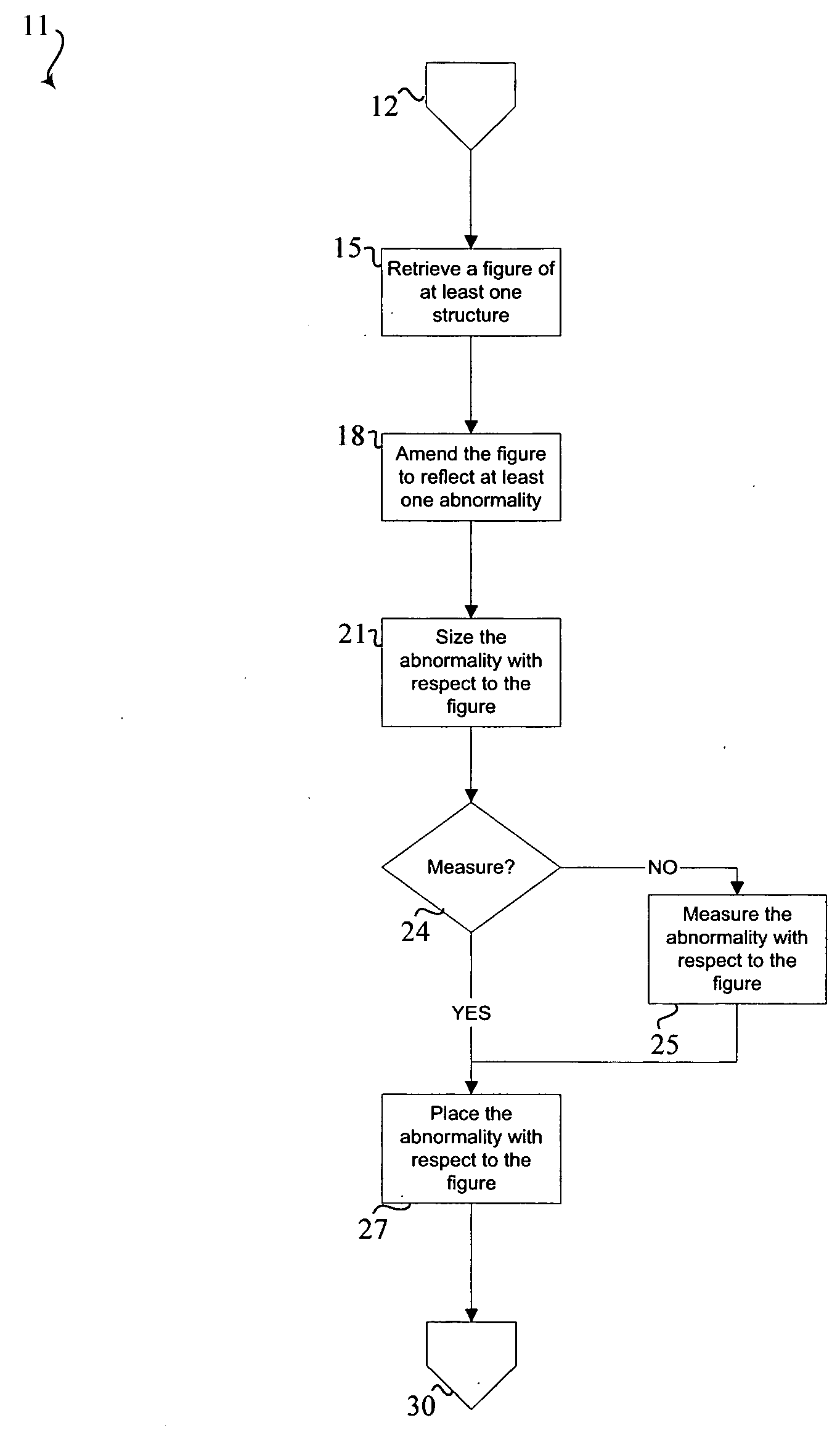
[0034] A method for generating a report of at least one abnormality evident on a medical image. The medical image includes at least a portion of at least one structure of the human body. The method includes retrieving a drawing of at least one structure. The retrieved drawing is amended to reflect the abnormality. The abnormality is sized and placed at a location on the retrieved drawing of the at least one structure, corresponding to the abnormalities location on the medical image. Placement of the sized abnormality generates an amended drawing representative of the medical image for inclusion in the report.

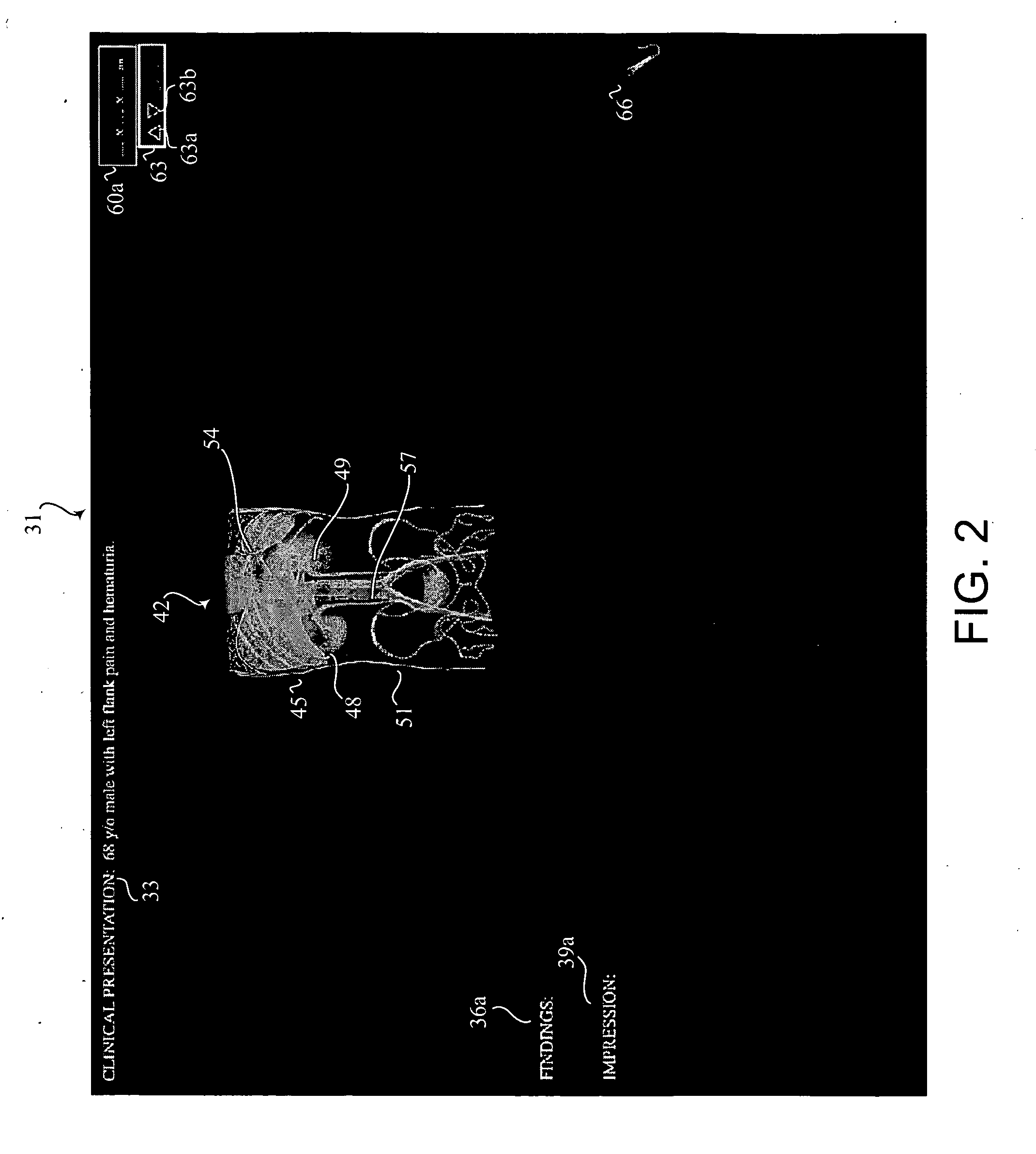
[0035] Within the teaching of this patent, the term “medical image” shall refer to any primary source of a healthcare practitioner's impressions. Under such a definition, both actual images from medical imaging devices (such as radiology films) or an examination of the actual patient the physician sees for physical exam are included. Thus, a general physician conducting a physi...


PUM


Abstract

A method and database for generating a report of findings of at least one abnormality evident in a medical image, the medical image including at least a portion of at least one structure of the body, the method includes retrieving a figure of the at least one structure. The retrieved figure is amended to reflect an abnormality. The abnormality is sized with respect to the figure of the at least one structure. The abnormality is placed at a location on the figure of the at least one structure according to an orientation of that abnormality in the medical image to generate an amended figure representative of the medical image for inclusion in the report.

Description

BACKGROUND OF THE INVENTION [0001] In current medical practice, diagnostic radiographic imaging examinations are usually ordered by clinical healthcare providers and interpreted by radiologists. A written interpretive report is generated by a radiologist and sent to the provider to convey and document the results of the examination. The interpreting radiologist, or other individual performing this function, will generally orally dictate a description and discussion of the abnormalities on the medical images, including text descriptive of his impression of the significance of those abnormalities. A transcriptionist or, in some instances, a computer with voice recognition software, will transform the spoken words of the radiologist into text. The radiologist dictates text documents to fulfill his professional responsibility and to convey the imaging examination results to the provider. [0002] The radiologist is tasked with the mental conversion of an image to text, the text being the ...

Claims


Application Information

Patent Timeline
Patent Type & Authority Applications(United States)
IPC IPC(8): G06F19/00G06K9/00
CPCG06F19/321G06Q50/24G06F19/3487G16H15/00G16H30/20
Inventor HEILBRUNN, KEN STEVENMILLER, GEORGE ANDREW
Owner ATLAS REPORTING
Why Eureka
Try Eureka