Virtual endoscopy

a virtual endoscope and endoscope technology, applied in the field of virtual endoscopes, can solve the problems of requiring heavy patient sedation, affecting the accuracy of endoscopes, and relatively invasive techniques, and achieve the disadvantage of requiring clinician input to identify the start voxel

Inactive Publication Date: 2008-05-22
TOSHIBA MEDICAL VISUALIZATION SYST EURO
View PDF13 Cites 26 Cited by
  • Summary
  • Abstract
  • Description
  • Claims
  • Application Information

AI Technical Summary

Benefits of technology

The technical effect of this patent is to create a more accurate dataset by eliminating off-centered artifacts such as artificial hip joints while still allowing for variations in coloπn position within a certain range.

Problems solved by technology

The technical problem addressed in this patent is how to accurately and efficiently identify the starting and endpoint positions when performing virtual endoscopy procedures on a biological object with a lumen. Existing methods involve either manual identification of these positions or computational intensity in determining them, leading to difficulties in optimizing the camera's movements during the procedure. There is a need for an improved automatic method for identifying these positions without compromising accuracy.

Method used


Image

  • Virtual endoscopy
  • Virtual endoscopy
  • Virtual endoscopy

Examples


Embodiment Construction

[0037]Virtual endoscopy is often used in the study of the colon. In this context virtual endoscopy is often referred to as virtual colonoscopy. Embodiments of the invention will hereafter be described by way of example in the specific context of virtual colonoscopy. However, it will be understood that embodiments of the invention may equally be employed in other applications of virtual endoscopy, and also in computer simulated fly-throughs of biological structures which are not normally the subject of conventional endoscopy procedures.

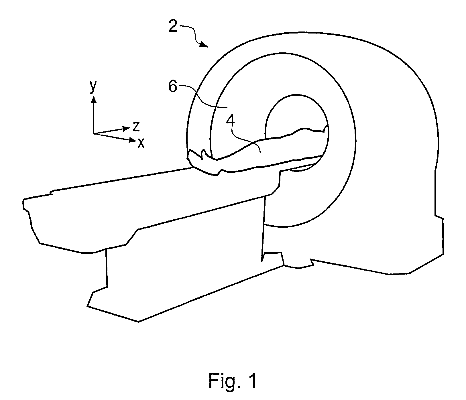
[0038]FIG. 1 is a schematic perspective view of a generic x-ray CT scanner 2 for obtaining a three-dimensional (3D) scan of a patient 4 to provide data suitable for virtual colonoscopy. The patient's abdominal region is placed within a circular opening 6 of the CT scanner 2 and a series of x-ray images are taken from directions around the patient. The raw image data are combined using tomographic techniques to produce a volume data set. The volume data...


PUM


Abstract

A method for automatically determining a start or finish location near to an end of a lumen in a medical image data set is described. The location may thus be used in determining a camera path for virtual endoscopy (e.g. colonoscopy). The data set comprises a plurality of voxels arranged along first, second and third directions and the method includes: segmenting the data set to identity a group of voxels classified as belonging to the lumen; selecting one of the axes of the data set as a primary direction based on an expected direction of the lumen at the end of interest; selecting a slice through the data set which is perpendicular to the primary direction and includes voxels at a spatial extremity of the group of voxels classified as belonging to the lumen along this direction; identifying a two-dimensional (2D) region of the voxels classified as belonging to the lumen within the selected slice; and selecting, based on the position of the 2D region, a position within the data set as the terminal location for the virtual colonoscopy camera path.

Description


Claims


Application Information

Patent Timeline
Owner TOSHIBA MEDICAL VISUALIZATION SYST EURO
Why Eureka
Try Eureka