Joint Detection and Localization of Multiple Anatomical Landmarks Through Learning

a technology of anatomical landmarks and joint detection, applied in image analysis, image enhancement, instruments, etc., can solve problems such as lack of generalization capability

Active Publication Date: 2009-02-05
SIEMENS HEALTHCARE GMBH
View PDF3 Cites 74 Cited by
  • Summary
  • Abstract
  • Description
  • Claims
  • Application Information

AI Technical Summary

Problems solved by technology

Although these methods achieve success in specific studies, they lack in generalization capability since their design incorporates the inherent structural information that needs to be detected.

Method used


Image

  • Joint Detection and Localization of Multiple Anatomical Landmarks Through Learning
  • Joint Detection and Localization of Multiple Anatomical Landmarks Through Learning
  • Joint Detection and Localization of Multiple Anatomical Landmarks Through Learning

Examples


Embodiment Construction

1. Introduction

[0030]As discussed in the Background section of this disclosure, landmark detection is a fundamental and important task in the area of medical image analysis. Reliable landmark detection paves the way to higher level analysis and understanding of medical images, e.g., segmentation, registration and image retrieval. Most of the existing studies address a specific landmark detection problem by carefully studying the appearance characteristics or shape priors of the landmarks. Although these methods achieve promising results in detecting specific landmarks, they lack the general potential to be extended to other studies.

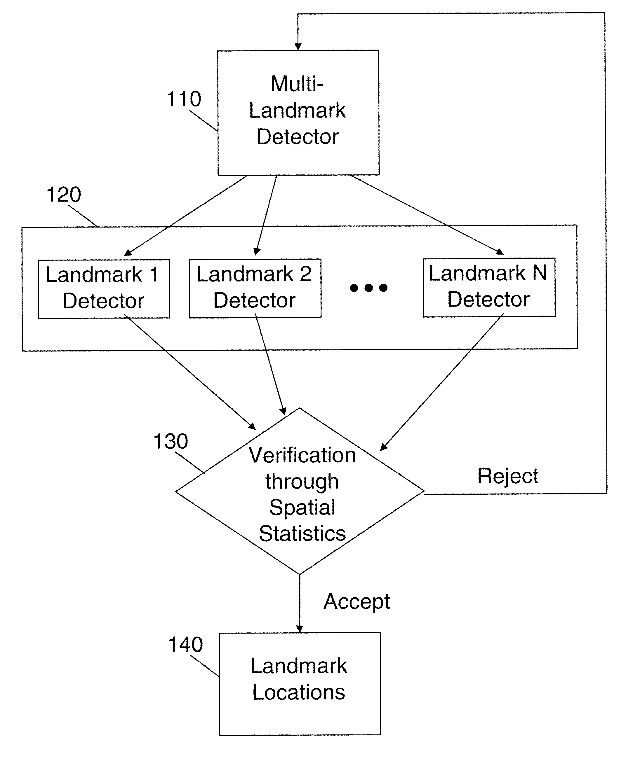
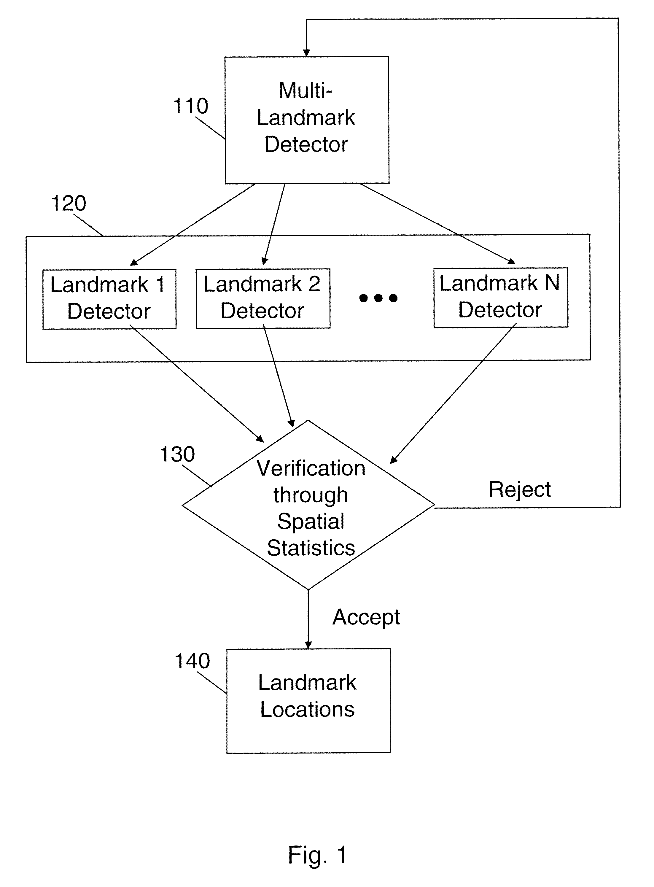
[0031]In the following, we present a learning-based method to detect and localize multiple anatomical landmarks from medical images, in accordance with an exemplary embodiment of the present invention. The method consists of three steps. First, a multi-landmark detector is applied to roughly detect candidate locations for multiple landmarks simultaneously...


PUM


Abstract

A method for detecting and localizing multiple anatomical landmarks in medical images including: receiving an input requesting identification of a plurality of anatomical landmarks in a medical image; applying a multi-landmark detector to the medical image to identify a plurality of candidate locations for each of the anatomical landmarks; for each of the anatomical landmarks, applying a landmark-specific detector to each of its candidate locations, wherein the landmark-specific detector assigns a score to each of the candidate locations, and wherein candidate locations having a score below a predetermined threshold are removed; applying spatial statistics to groups of the remaining candidate locations to determine, for each of the anatomical landmarks, the candidate location that most accurately identifies the anatomical landmark; and for each of the anatomical landmarks, outputting the candidate location that most accurately identifies the anatomical landmark.

Description

CROSS-REFERENCE TO RELATED APPLICATION[0001]This application claims the benefit of U.S. Provisional Application No. 60 / 953,553, filed Aug. 2, 2007, the disclosure of which is incorporated by reference herein in its entirety.BACKGROUND OF THE INVENTION[0002]1. Technical Field[0003]The present invention relates to landmark detection in medical images.[0004]2. Discussion of the Related Art[0005]In the area of medical image analysis, anatomical landmark detection often plays a fundamental and critical role. High level image analysis and understanding, e.g., segmentation [M. Rousson, A. Khamene, M. Diallo, J. C. Celi, and F. Sauer, “Constrained surface evolutions for prostate and bladder segmentation in ct images”, in Computer Vision for Biomedical Image Applications, pp. 251-260, 2005], registration [Y. Zhan, Y. Ou, M. Feldman, J. Tomaszeweski, C. Davatzikos, and D. Shen, “Registering histologic and mr images of prostate for image-based cancer detection”, Academic Radiology, 14, pp. 136...

Claims


Application Information

Patent Timeline
Patent Type & Authority Applications(United States)
IPC IPC(8): G06K9/00
CPCG06T7/0044G06T2207/30004G06T2207/10072G06T7/74
Inventor DIKMEN, MERTZHAN, YIQIANGZHOU, XIANG
Owner SIEMENS HEALTHCARE GMBH
Why Eureka
Try Eureka