Method for forming organ
a technology for organs and tissues, applied in the field of organ formation, can solve the problems of undifferentiated cells, complex process for organ formation,
- Summary
- Abstract
- Description
- Claims
- Application Information
AI Technical Summary
Problems solved by technology
Method used
Image
Examples
example 1
Formation of Heart Using RXR Ligand
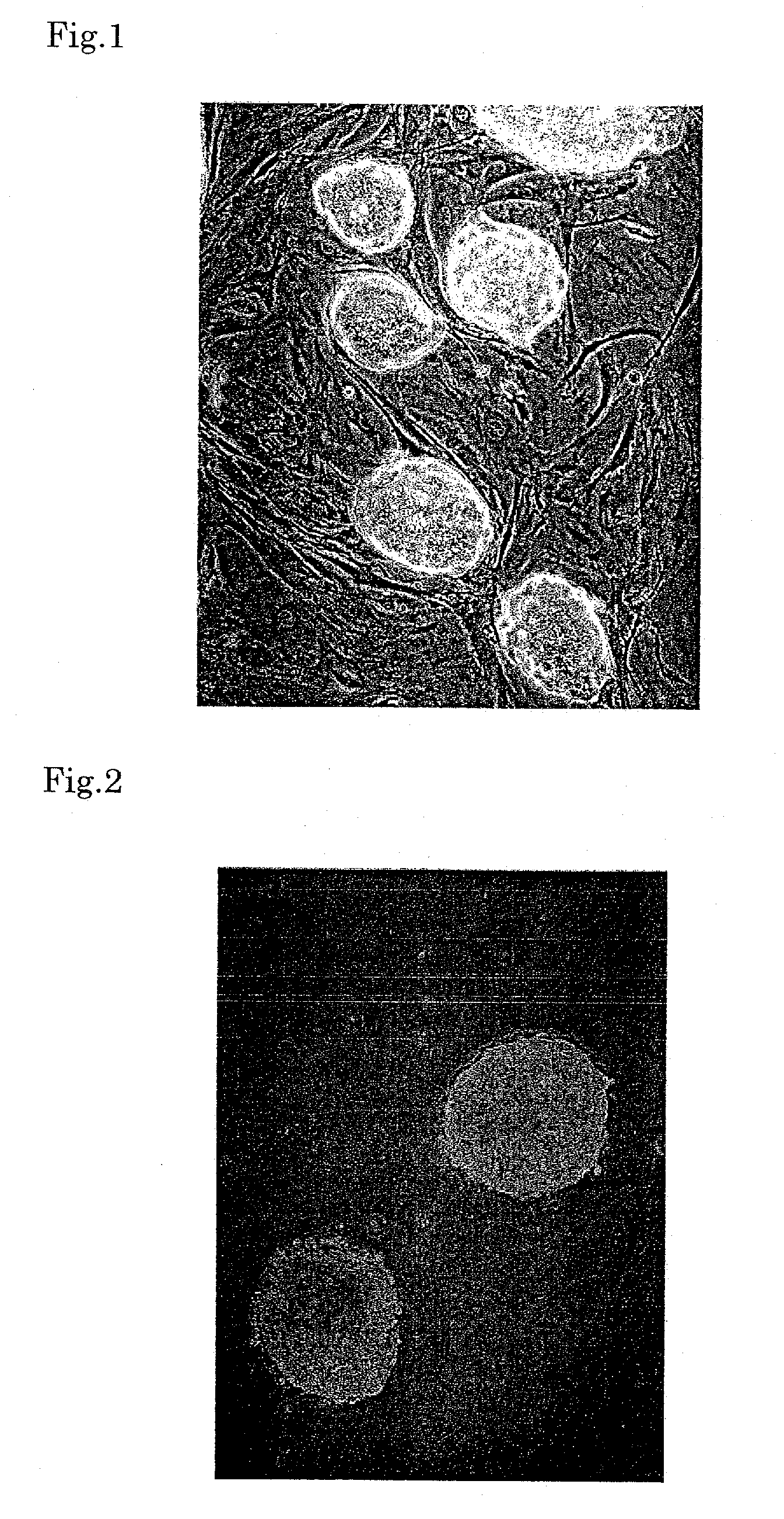
[0061]As ES cells, E 14 cells of the 29 mouse strain (ATCC #: CRL-1821) or B6 cells of the C57BL mouse strain (ATCC #: SCRC-1002) were used. Mouse fetal fibroblasts prepared from a mouse embryo on the 13th day were inoculated on a 0.1% gelatin-coated culture dish, and cultured in a medium containing 12% fetal bovine serum (Gibco), 100 U / ml penicillin (Sigma) and 100 μg / ml streptomycin (Sigma) for 24 hours. These cells were treated with 10 μg / ml mytomicin C (MMC, Sigma) for four hours to inhibit cell division and then washed twice with phosphate-buffered saline (PBS) to remove MMC. The ES cells were inoculated on these fibroblasts (feeder). As the medium for the ES cells, D-MEM (high glucose, Gibco) containing 15% fetal bovine serum for ES (Gibco), 2 mM L-glutamine (Gibco), MEM non-essential amino acid (Sigma), 1 mM sodium pyruvate (Gibco), 0.0007% β-mercaptoethanol (Sigma), 1000 U / ml Leukemia inhibiting factor (Chemicon), 100 U / ml penicillin (Sigma...
example 2
Formation of Smooth Muscle and Adipocytes Using RXR Ligand
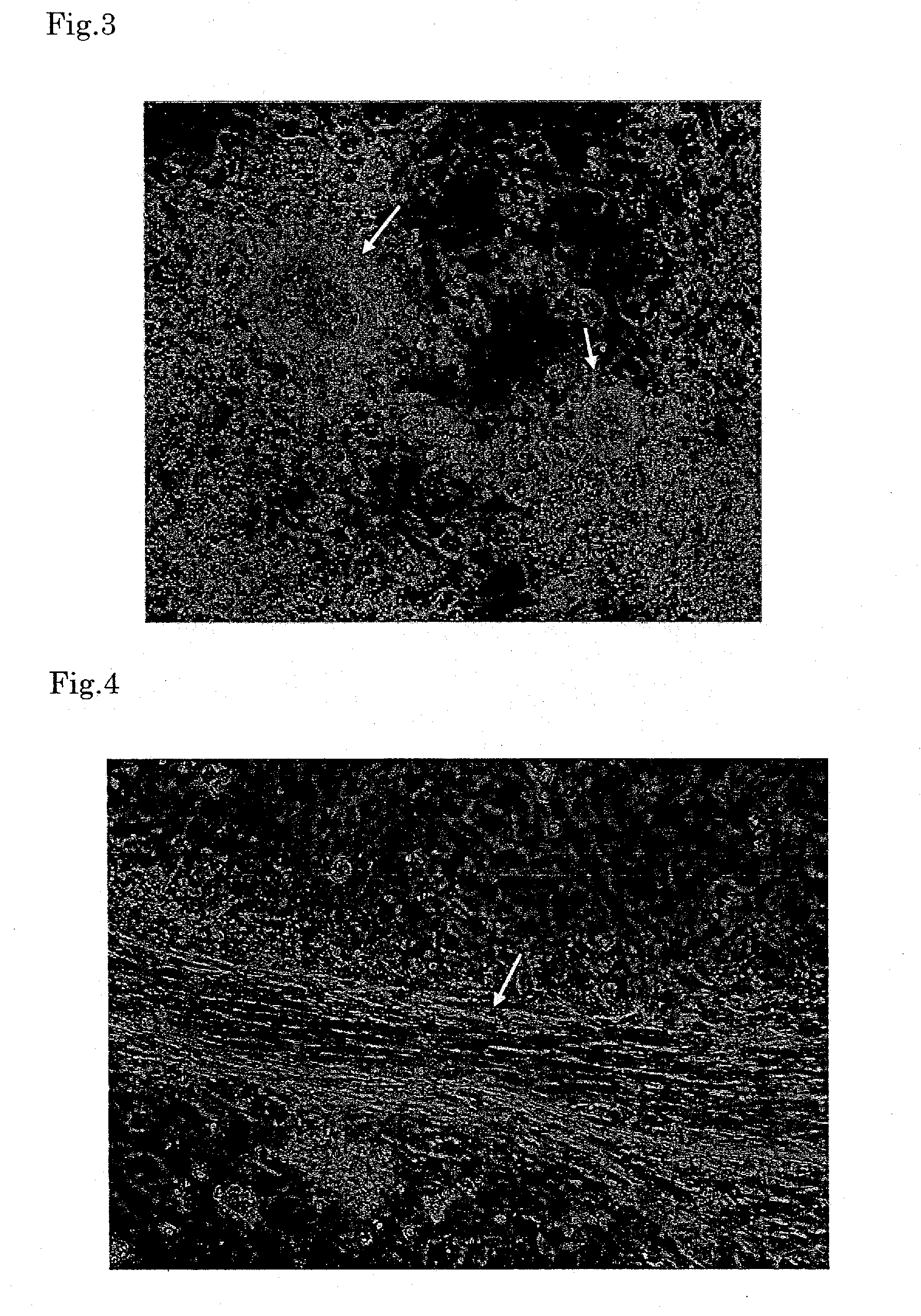
[0064]When the embryoid bodies were treated with 1×10⁻⁵ to 2×10⁻⁶ M PA024 and then further cultured in the same manner as in Example 1, smooth muscle-like cells were formed in about one to two weeks. The resulting cell aggregate showed slow peristalsis. The frequency of appearance was 0.1 to 0.2 cell aggregate per embryoid body (FIG. 4). Further, appearance of adipocytes was observed in about two weeks, and after the culture was continued for 20 days, about 30 to 40% of the total cells became adipocytes, which was substantially 100% in terms of the value per embryoid body. In comparison with the control group (treated with 1×10¹⁵ M all-trans retinoic acid), two or three times more adipocytes were formed in the PA024 treated group (FIG. 5).
example 3
Formation of Pancreas Using Combination of Am80 as RAR Ligand and Activin
[0065]Primary mouse embryonic fibroblasts (PMEFs), of which cell division was inhibited by treatment with Mytomicin-C (10 μg / ml, 3 hours), were inoculated on a 10-cm culture dish (TPP) coated overnight with 0.1% gelatin, cultured for more than one day and used as a feeder layer. The ES-E14TG2a cells (ATCC # CRL-1821), which are widely used as mouse ES cells, were inoculated on the feeder layer and cultured. As the medium for ES cells, DMEM (high glucose, Gibco) containing 15% fetal bovine serum (FBS, Gibco), non-essential amino acid, 0.007%, mercaptoethanol, 1000 U / ml Leukemia inhibiting factor (Chemicon), 100 U / ml penicillin, and 0.1 mg / ml streptomycin was used. The medium was exchanged every day.
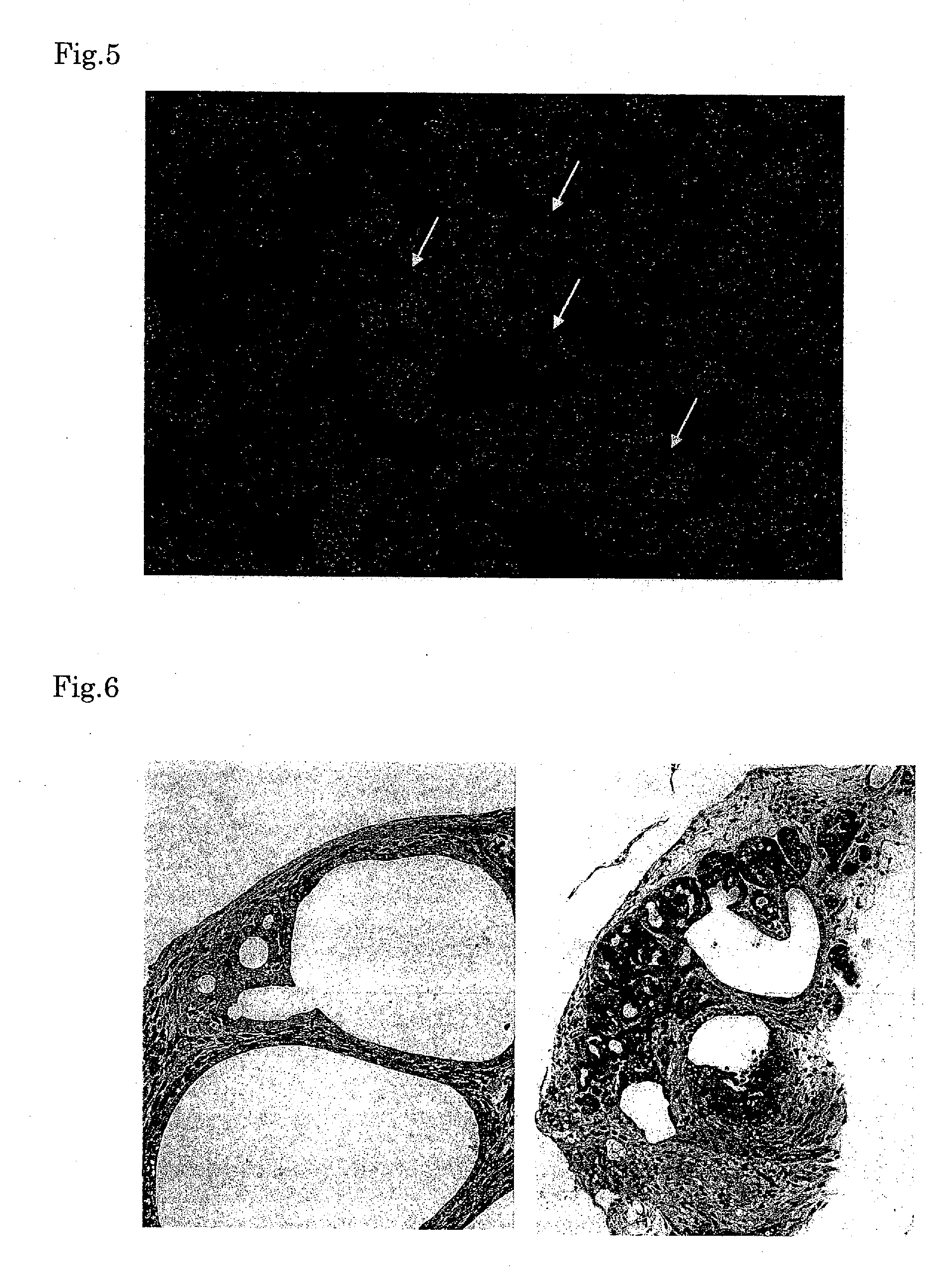
[0066]EBs were formed from colonies 72 hours after the inoculation of the ES cells. ES cell colonies were washed once with Dulbecco's PBS, then added with 1 ml of 1 mg / ml collagenase / dispase (Roche) per 10-cm dish and...
PUM
| Property | Measurement | Unit |
|---|---|---|
| concentration | aaaaa | aaaaa |
| humidity | aaaaa | aaaaa |
| humidity | aaaaa | aaaaa |
Abstract
Description
Claims
Application Information
 - R&D
- Intellectual Property
- Life Sciences
- Materials
- Tech Scout
- Unparalleled Data Quality
- Higher Quality Content
- 60% Fewer Hallucinations
Browse by: Latest US Patents, China's latest patents, Technical Efficacy Thesaurus, Application Domain, Technology Topic, Popular Technical Reports.
© 2025 PatSnap. All rights reserved.Legal|Privacy policy|Modern Slavery Act Transparency Statement|Sitemap|About US| Contact US: help@patsnap.com


