
Identification of regions of interest and extraction of time value curves in imaging procedures

a time value curve and imaging procedure technology, applied in the field of identification, can solve the problems of difficult to draw difficult to determine the size, shape and location of the regions of interest, and requires valuable operator tim

Inactive Publication Date: 2009-12-24
BAYER HEALTHCARE LLC
View PDF99 Cites 21 Cited by
  • Summary
  • Abstract
  • Description
  • Claims
  • Application Information

AI Technical Summary

Benefits of technology

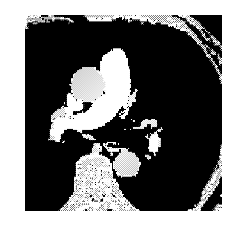
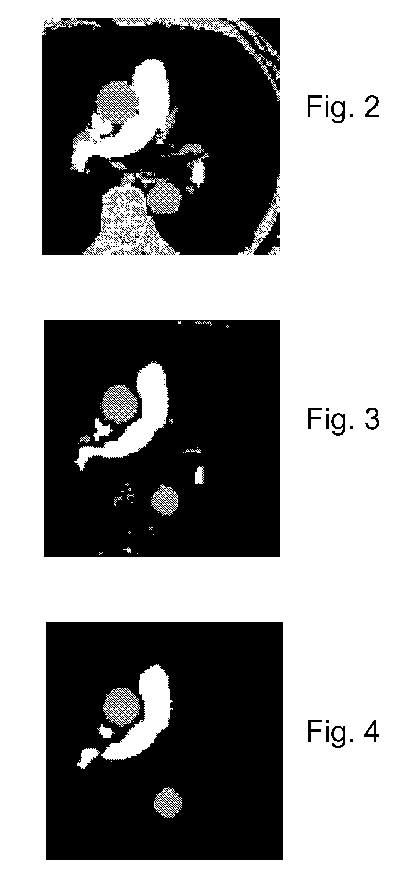
[0010]The segments can, for example, be computed using a clustering technique such as K-means clustering. K clusters can, for example, be computed by minimizing the sum of squares of distances between data points and K cluster centroids, wherein K is an integer corresponding to the predetermined number of correlated segments.
[0016]The method can further include filtering the segmented image to eliminate pixels that are not well correlated with neighboring pixels. Each pixel of the segmented image can, for example, be compared with all eight of its neighboring pixels in filtering.

Problems solved by technology

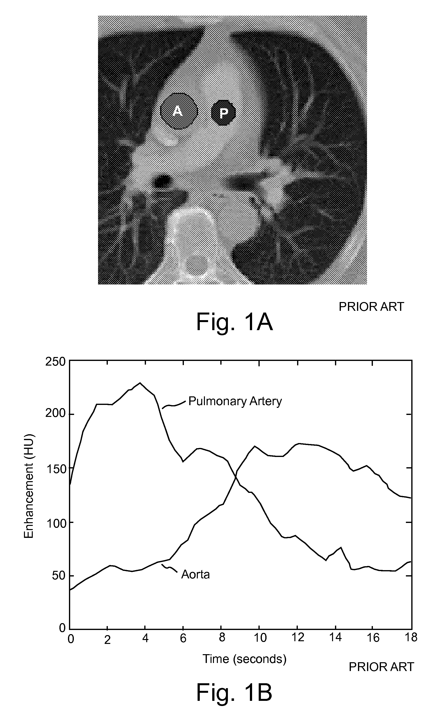
Determining the size, shape, and location of the regions of interest is challenging and requires valuable operator time (for example, to go to a separate evaluation screen, to load the images, to draw the region(s) of interest, and to use the resulting data to generate and / or adjust the contrast / scan protocol).
It can be particularly difficult to draw the regions of interest on the image before contrast has been injected.
Moreover, factors such as patient movement can result in substantial inaccuracies.

Method used


Image

  • Identification of regions of interest and extraction of time value curves in imaging procedures
  • Identification of regions of interest and extraction of time value curves in imaging procedures
  • Identification of regions of interest and extraction of time value curves in imaging procedures

Examples


Embodiment Construction

[0036]In several embodiments of the present invention, one or more regions of interest are determined in a portion of a patient's body by the devices, systems and / or methods of the present invention via analysis of a plurality of datasets of values (for example, pixel values) of the portion of the patient's body (in a time series of such datasets) without the requirement that an operator manually draw or otherwise define the regions of interest. The dataset of values (for example, point values such as pixel intensity values) need not be displayed to an operator as a viewed image during determination of the one or more regions of interest. In a number of embodiments, after one or more regions of interest are determined, time value curves (for example, time enhancement curves in the case that a contrast enhancement medium is injected) for the regions of interest are computed from the dataset series (for example, a contrast-enhanced transit bolus image series).

[0037]As used herein, the...


PUM


Abstract

A method of extracting at least one time-value curve to determine a protocol in an imaging procedure using an imaging system, includes: determining a first N-dimensional data set of pixel values of a portion of a body of the patient at a first time using the imaging system, wherein N is an integer; determining at least a second N-dimensional data set of pixel values of the portion at a second time using the imaging system; computing a predetermined number of correlated segments of the imaged portion corresponding to a predetermined number of regions of interest of the patient by computing a similarity metric of a time series of pixel values; computing the at least one time-value curve for at least one of the regions of interest; and determining a protocol for a diagnostic scan using the image system based at least in part upon data from the time value curve.

Description

BACKGROUND OF THE INVENTION[0001]The present invention relates to identification of one or more regions of interest in imaging procedures and, particularly, to identification of one or more regions of interest with a minimum of or without manual intervention and subsequent extraction of time value curves for the one or more regions of interest.[0002]The following information is provided to assist the reader to understand the invention disclosed below and the environment in which it will typically be used. The terms used herein are not intended to be limited to any particular narrow interpretation unless clearly stated otherwise in this document. References set forth herein may facilitate understanding of the present invention or the background of the present invention. The disclosure of all references cited herein are incorporated by reference.[0003]Image enhancement via injection of contrast enhancement media or contrast media in imaging procedures (for example, computed tomography...

Claims


Application Information

Patent Timeline
Patent Type & Authority Applications(United States)
IPC IPC(8): G06K9/00G06K9/34
CPCG06K9/6223G06K2209/051G06T2207/30061G06T7/0087G06T2207/10081G06T7/0081G06T7/0012G06T7/0016G06T7/11G06T7/143G06V2201/031G06V10/763G06F18/23213
Inventor KEMPER, COREYKALAFUT, JOHN F.
Owner BAYER HEALTHCARE LLC
Learn More