Method and system for delineation of vasculature

a vasculature and vasculature technology, applied in the field of medical imaging examination methods and apparatuses, can solve the problems of reducing the clinical value of vasculature,

Active Publication Date: 2010-03-04
GENERAL ELECTRIC CO
View PDF3 Cites 45 Cited by
  • Summary
  • Abstract
  • Description
  • Claims
  • Application Information

AI Technical Summary

Benefits of technology

The patent describes a method for extracting the vasculature in the lung fields by using filters based on patient parameters. This helps to suppress the soft tissue in the lung fields and improve the accuracy of the extraction process. The technical effect of this method is to provide a more reliable and accurate way of extracting the vasculature in the lung fields, which can be useful in various medical applications such as diagnostics and treatment planning.

Problems solved by technology

The technical problem addressed in this patent text is the need for a system that can delineate the vasculature in anatomical regions, such as the lungs, from the surrounding background using digital X-ray image data. This is important for improving the detection of disease states in the vasculature and simplifying the clinical workflow of the diagnostic imaging system. The challenge is that traditional X-ray radiographs have low sensitivity and selectivity for extracting vascular tree structures, which can lead to missed detection of disease states and misdiagnosis.

Method used


Image

  • Method and system for delineation of vasculature
  • Method and system for delineation of vasculature
  • Method and system for delineation of vasculature

Examples


Embodiment Construction

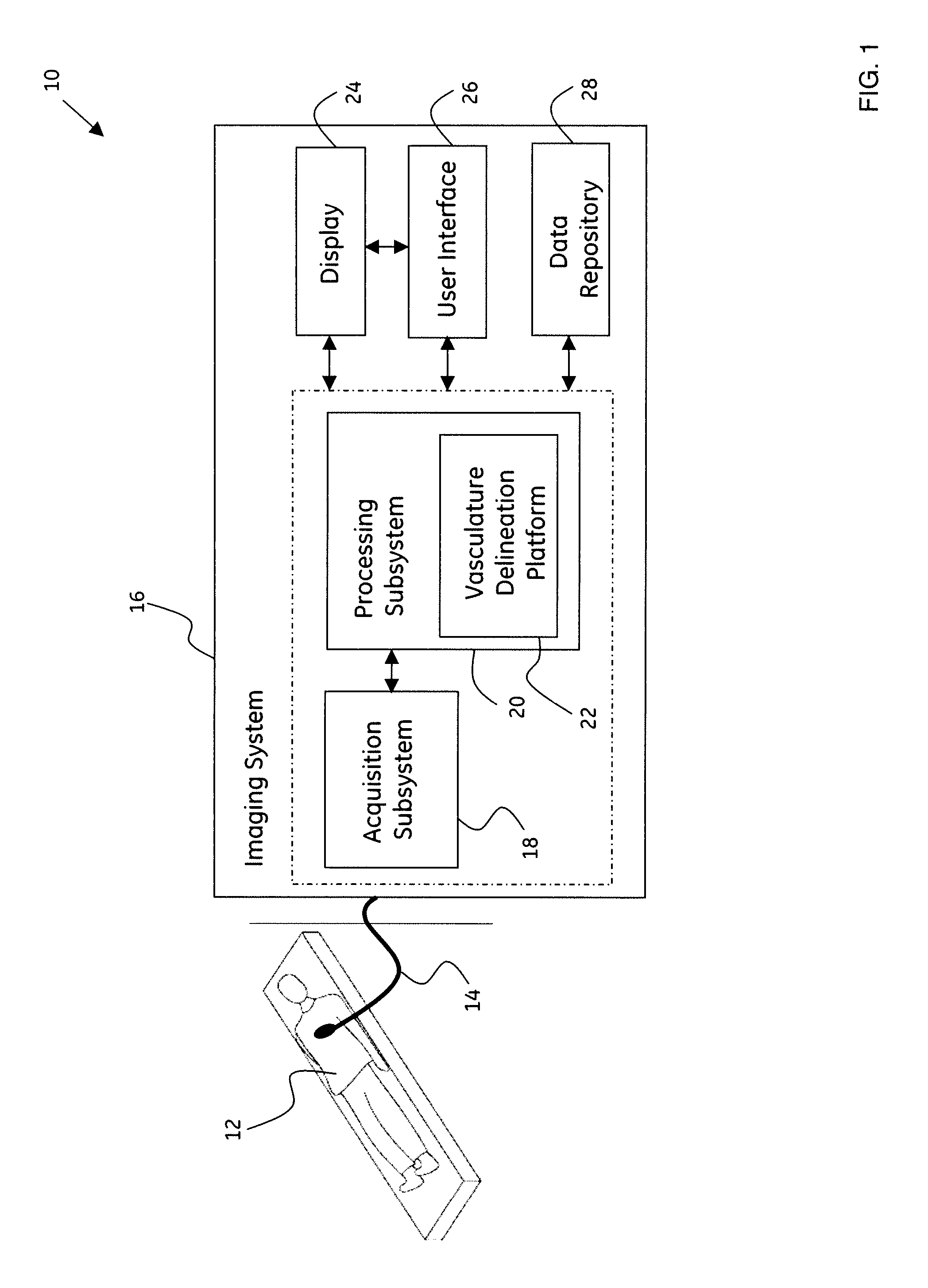
[0024]As will be described in detail hereinafter, a method for automatically delineating image data representative of vasculature in an anatomical region and a system for automatically delineating image data representative of vasculature in the anatomical region configured to optimize detection of disease in the vasculature and simplify clinical workflow in a diagnostic imaging system, are presented. Employing the method and system described hereinafter, the system for the delineation of image data representative of vasculature may be configured to facilitate substantially superior detection of disease in the vasculature, thereby simplifying the clinical workflow of the detection of disease states.

[0025]Although, the exemplary embodiments illustrated hereinafter are described in the context of a medical imaging system, it will be appreciated that use of the diagnostic system in industrial applications are also contemplated in conjunction with the present technique.

[0026]FIG. 1 is a ...


PUM


Abstract

A method for automatically delineating image data corresponding to vasculature in an anatomical region is presented. The method includes obtaining image data corresponding to soft tissue representative of the anatomical region. In addition, the method includes segmenting image data corresponding to a region of interest from image data corresponding to the anatomical region based on a patient population to obtain image data corresponding to a segmented region of interest. Furthermore, the method includes extracting image data corresponding to vasculature associated with the segmented region of interest by suppressing image data corresponding to soft tissue representative of the segmented region of interest. Systems and computer-readable medium that afford functionality of the type defined by this method are also contemplated in conjunction with the present technique.

Description


Claims


Application Information

Patent Timeline
Owner GENERAL ELECTRIC CO
Why Eureka
Try Eureka