Apparatus and method for providing a dynamic 3D ultrasound image

a technology of ultrasound image and apparatus, applied in the field of apparatus for capturing and reconstructing a three-dimensional (3d) ultrasound image of a vessel, can solve the problem of insufficient information obtained from the ultrasound imag

Inactive Publication Date: 2010-07-08
IND TECH RES INST
View PDF8 Cites 6 Cited by
  • Summary
  • Abstract
  • Description
  • Claims
  • Application Information

AI Technical Summary

Problems solved by technology

However, if 2D ultrasonic images are used to be the bases for further diagnoses, the information obtained therefrom seem insufficient therefor since the power of ultrasonic waves will apperently lose while those ultrasonic waves meet the calcified occlusion.

Method used


Image

  • Apparatus and method for providing a dynamic 3D ultrasound image
  • Apparatus and method for providing a dynamic 3D ultrasound image
  • Apparatus and method for providing a dynamic 3D ultrasound image

Examples


Embodiment Construction

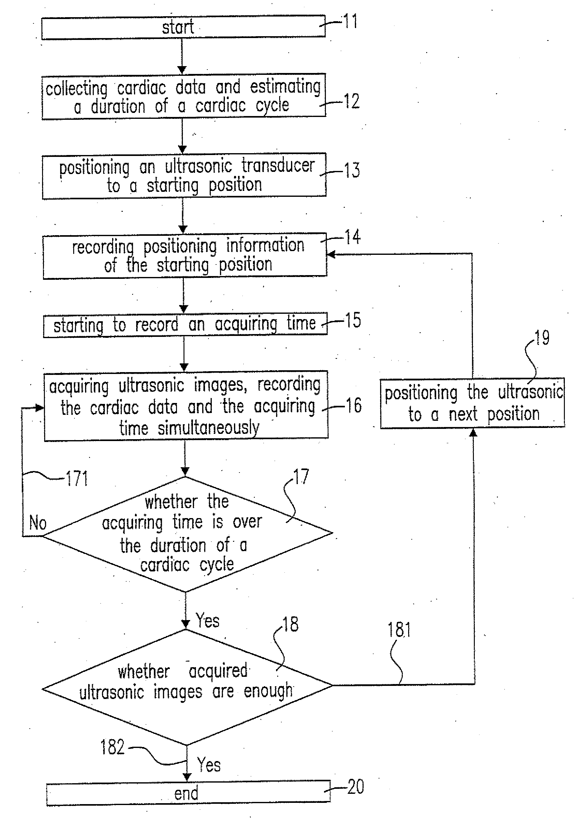
[0021]Please refer to FIG. 4(A), which is a flow chart illustrating the method of the present disclosure for acquiring the ultrasonic images. When start to acquire ultrasonic images (Step 11), a duration of one cardiac cycle of a subject is obtained by the subject's cardiac data, e.g. an electrocardiogram (Step 12). Then, an ultrasonic transducer will be posited to a starting position of target region on the shin (Step 13), where the positing process can be driven by an auxiliary system such as a positioning system having an electronic motor and positioning information of the starting position is recorded (Step 14), and the target region is typically a vessel or an organ. After the ultrasonic transducer is well posited, an acquiring time, i.e. the time for acquiring ultrasonic image, starts to be recorded (Step 14), meanwhile, the ultrasonic transducer is triggered to acquire ultrasonic image (Step 15). By the ultrasonic transducer, the ultrasonic images at the start position will c...


PUM


Abstract

A system and method thereof for capturing and reconstructing a dynamic 3D ultrasound image of a blood vessel are provided. The present system includes a ultrasound transducer, a motor positioning system, an electrocardiograph, a microprocessor for processing ultrasound images and signals capturing by the ultrasound transducer, and a monitor displaying the dynamic 3D ultrasound image. The motor positioning system controls the ultrasound transducer to capture 2D ultrasound images of the blood vessel at several locations in a predetermined urea. During the capturing process, capturing times, electrocardiograms and 3D locations of those captured 2D ultrasound images are simultaneously recoded. Finally, those captured 2D ultrasound images are reconstructed to the dynamic 3D ultrasound image of the blood vessel according to the time bases based on the phases of the electrocardiograms.

Description

TECHNICAL FIELD[0001]The present disclosure relates to an apparatus for capturing and reconstructing a three-dimensional (3D) ultrasound image of a vessel and the method thereof. More particularly, the present apparatus and method are used for imaging and displaying a dynamic 3D ultrasound image of the vessel.DESCRIPTION OF RELATED ART[0002]The ultrasonic detection is popular in medical procedure since its characteristics of cheapness and noninvasion. Presently, the ultrasonic detection is performed via repeatedly scanning a specific region of the patient with the handheld ultrasonic transducer. During the ultrasonic detection, the piezoelectric transducer of the ultrasonic transducer generates ultrasonic waves with 2 to 13 MHz to introduce into the patient's body. Then, the oscillation of the ultrasonic transducer receives the ultrasonic echoes reflexed from the interfaces of various tissues and transforms thereto electronic pulses. The electronic pulses are sequentially transmitte...

Claims


Application Information

Patent Timeline
Patent Type & Authority Applications(United States)
IPC IPC(8): A61B8/14A61B5/0402
CPCA61B5/02007A61B5/201A61B8/13A61B8/543A61B8/4254A61B8/483A61B8/145
Inventor LIN, CHIN-ANWU, GUO-ZUAPAO, SUN-HUAHUANG, CHIEN-HSINGYU, HSING-CHENGCHEN, HUNG-YUEH
Owner IND TECH RES INST
Why Eureka
Try Eureka