Devices and methods for treating tissue

a technology for treating tissue and devices, applied in the field of devices and methods for treating tissue, can solve the problems of difficult to modify certain tissues to achieve a desired effect, more technically challenging procedures, and reduced or even no direct visualization

Active Publication Date: 2011-03-10
SPINAL ELEMENTS INC
View PDF65 Cites 100 Cited by
  • Summary
  • Abstract
  • Description
  • Claims
  • Application Information

AI Technical Summary

Problems solved by technology

Minimally invasive (or “less invasive”) surgical procedures often involve modifying tissue through one or more small incisions or percutaneous access, and thus may be more technically challenging procedures.
Some of the challenges of minimally invasive tissue modification procedures include working in a smaller operating field, working with smaller devices, and trying to operate with reduced or even no direct visualization of the tissue (or tissues) being modified.
For example, using arthroscopic surgical techniques for repairing joints such as the knee or the shoulder, it may be quite challenging to modify certain tissues to achieve a desired result, due to the required small size of arthroscopic instruments, the confined surgical space of the joint, lack of direct visualization of the surgical space, and the like.
It may be particularly challenging in some surgical procedures, for example, to cut or contour bone or ligamentous tissue with currently available minimally invasive tools and techniques.
For example, trying to shave a thin slice of bone off a curved bony surface, using a small-diameter tool in a confined space with little or no ability to see the surface being cut, as may be required in some procedures, may be incredibly challenging or even impossible using currently available devices.
The most common form of spinal stenosis occurs in the lower (or lumbar) spine and can cause severe pain, numbness and / or loss of function in the lower back and / or one or both lower limb.
Impingement of neural and / or neurovascular tissue in the spine by one or more of these tissues may cause pain, numbness and / or loss of strength or mobility in one or both of a patient's lower limbs and / or of the patient's back.
When these conservative treatment options fail and symptoms are severe, as is frequently the case, surgery may be required to remove impinging tissue and decompress the impinged nerve tissue.
Removal of vertebral bone, as occurs in laminectomy and facetectomy, often leaves the affected area of the spine very unstable, leading to a need for an additional highly invasive fusion procedure that puts extra demands on the patient's vertebrae and limits the patient's ability to move.
Unfortunately, a surgical spine fusion results in a loss of ability to move the fused section of the back, diminishing the patient's range of motion and causing stress on the discs and facet joints of adjacent vertebral segments.
Such stress on adjacent vertebrae often leads to further dysfunction of the spine, back pain, lower leg weakness or pain, and / or other symptoms.
Furthermore, using current surgical techniques, gaining sufficient access to the spine to perform a laminectomy, facetectomy and spinal fusion requires dissecting through a wide incision on the back and typically causes extensive muscle damage, leading to significant post-operative pain and lengthy rehabilitation.
Thus, while laminectomy, facetectomy, and spinal fusion frequently improve symptoms of neural and neurovascular impingement in the short term, these procedures are highly invasive, diminish spinal function, drastically disrupt normal anatomy, and increase long-term morbidity above levels seen in untreated patients.

Method used


Image

  • Devices and methods for treating tissue
  • Devices and methods for treating tissue
  • Devices and methods for treating tissue

Examples


Embodiment Construction

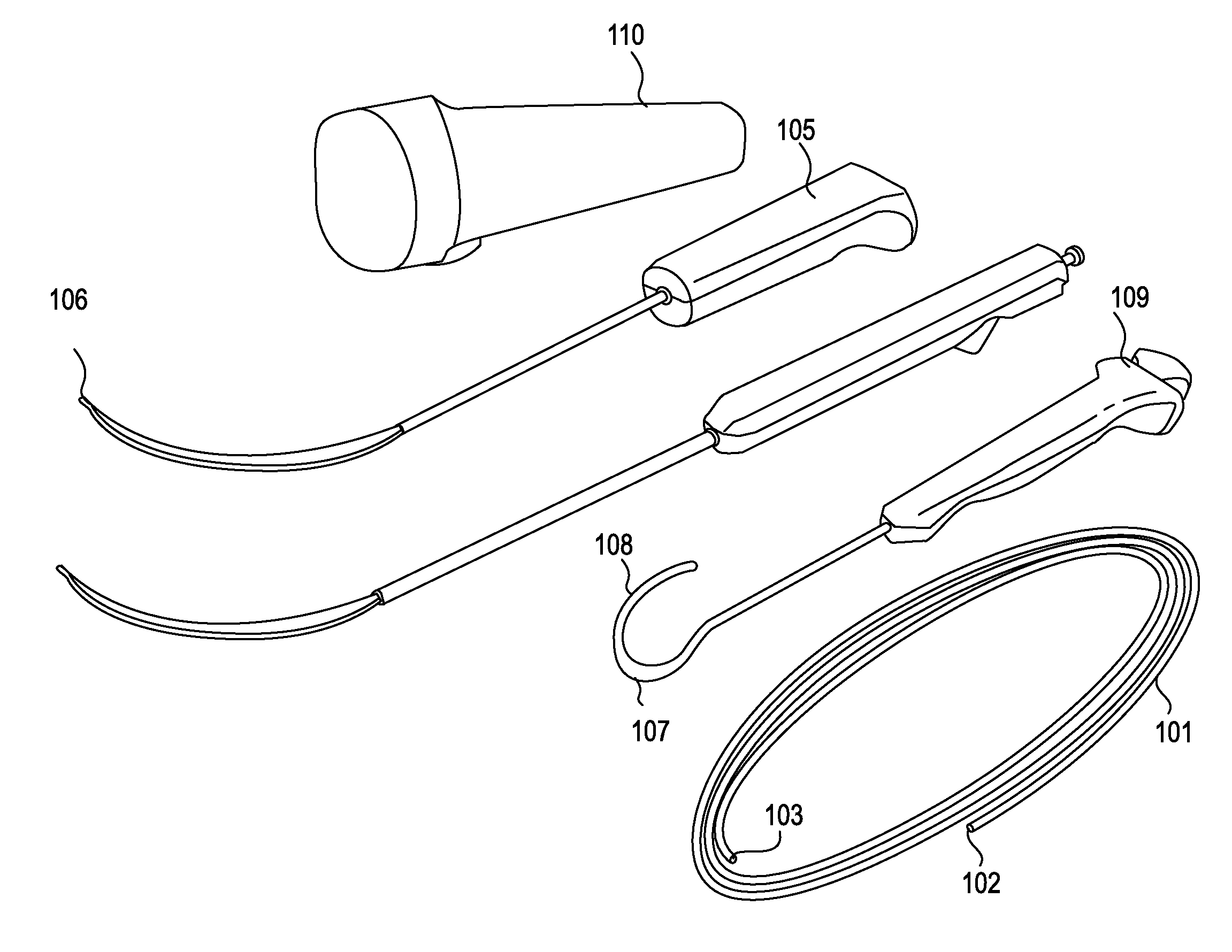
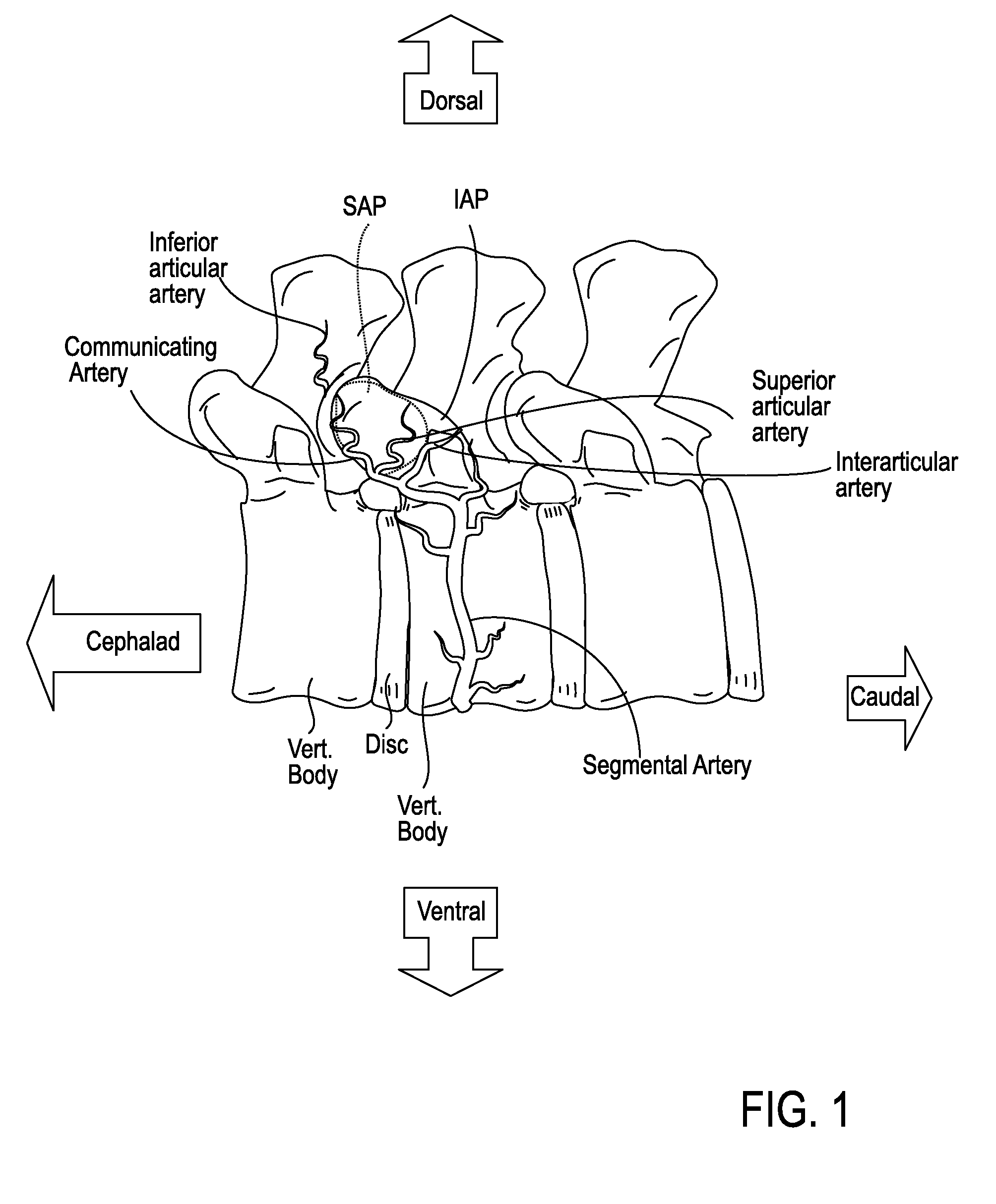
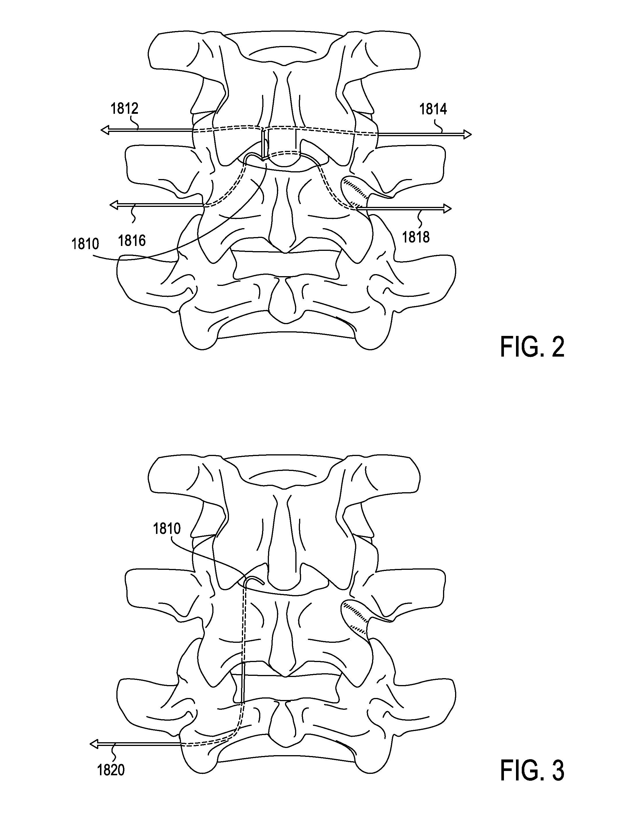
[0050]The devices, systems and methods described herein may be use in any appropriate surgical procedure, particularly for the surgical treatment of spinal stenosis. For example, described herein are systems including one or more of the following devices: a guidewire, a probe for positioning a guidewire, and a tissue modification device for use with the guidewire. As described herein, the systems and methods may be used to decompress one or more spinal regions. In particular, any of these devices may be used to decompress nerve roots within the spinal anatomy along various paths. Because these devices are flexible, and may be appropriately sized and shaped to fit within a neural foramen, these devices may be used to accesses appropriate regions of the spine from a single access point (e.g., from the patient's midline or near-midline region). While the systems and methods described herein may used to decompress one or more spinal regions they may also be configured to avoid portions ...


PUM


Abstract

Described herein are devices, systems and methods for treating target tissue in a patient's spine. In general, the methods include the steps of advancing a wire into the patient from a first location, through a neural foramen, and out of the patient from a second location; connecting a tissue modification device to the wire; positioning the tissue modification device through the neural foramen using the wire; modifying target tissue in the spine by moving the tissue modification device against the target tissue; and delivering an agent to modified target tissue, wherein the agent is configured to inhibit blood flow from the modified target tissue. In some embodiments, the step of modifying target tissue comprises removing target tissue located ventral to the superior articular process while avoiding non-target tissue located lateral to the superior articular process.

Description

CROSS REFERENCE TO RELATED APPLICATIONS[0001]This patent application is a continuation-in-part to U.S. patent application Ser. No. 11 / 251,205, titled “DEVICES AND METHODS FOR TISSUE ACCESS”, filed on Oct. 15, 2005; which claims the benefit of U.S. Provisional Patent Application No. 60 / 619,306, filed Oct. 15, 2004, U.S. Provisional Patent Application No. 60 / 622,865, filed Oct. 28, 2004, U.S. Provisional Patent Application No. 60 / 681,719, filed May 16, 2005, U.S. Provisional Patent Application No. 60 / 681,864, filed May 16, 2005, and U.S. Provisional Patent Application No. 60 / 685,190, filed May 27, 2005, each of which is incorporated by reference herein in its entirety.[0002]This patent application is also a continuation-in-part to U.S. patent application Ser. No. 11 / 468,247, titled “TISSUE ACCESS GUIDEWIRE SYSTEM AND METHOD”, filed on Aug. 29, 2006. This application is herein incorporated by reference in its entirety.[0003]This patent application also claims priority to U.S. Provision...

Claims


Application Information

Patent Timeline
Patent Type & Authority Applications(United States)
IPC IPC(8): A61M25/00A61B17/16
CPCA61B17/1659A61B2017/145A61B17/320016A61B17/3401A61B17/3403A61B17/3468A61B18/1477A61B18/1487A61B19/40A61B2017/00261A61B2017/003A61B2017/00331A61B2017/00867A61B2017/143A61B2017/22038A61B2017/22049A61B2017/320004A61B2017/320044A61B2017/32006A61B2018/1407A61B2018/1425A61B2019/461A61B2019/481A61F2/0045A61N1/0551A61N1/36017A61B17/1671A61B90/04A61B2090/061A61B2090/08021A61B17/149A61B17/32A61M25/0067A61M25/09A61M2210/1003
Inventor WALLACE, MICHAEL P.GARABEDIAN, ROBERTARCENIO, GREGORY B.BLEICH, JEFFERY L.SAADAT, VAHIDCHUNG, WINNIE
Owner SPINAL ELEMENTS INC
Why Eureka
Try Eureka