Set of oligonucleotide probes as well as methods and uses thereto
a technology of oligonucleotide probes and probes, applied in the field of oligonucleotide probes, can solve the problems of inability to preserve the slides for storage and review, high cost of each test, and longer time required for slide scoring
- Summary
- Abstract
- Description
- Claims
- Application Information
AI Technical Summary
Benefits of technology
Problems solved by technology
Method used
Image

Examples
example 1
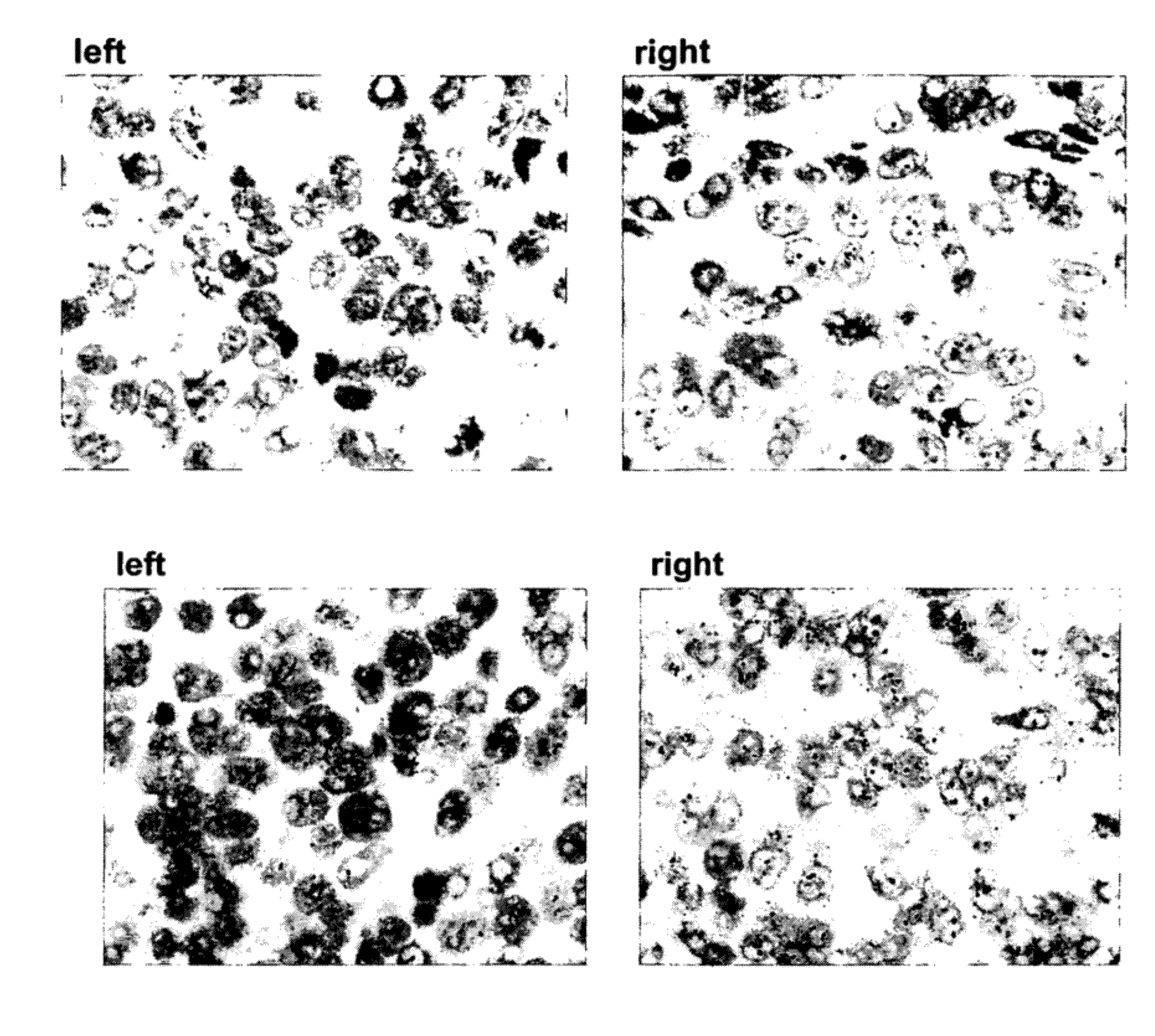
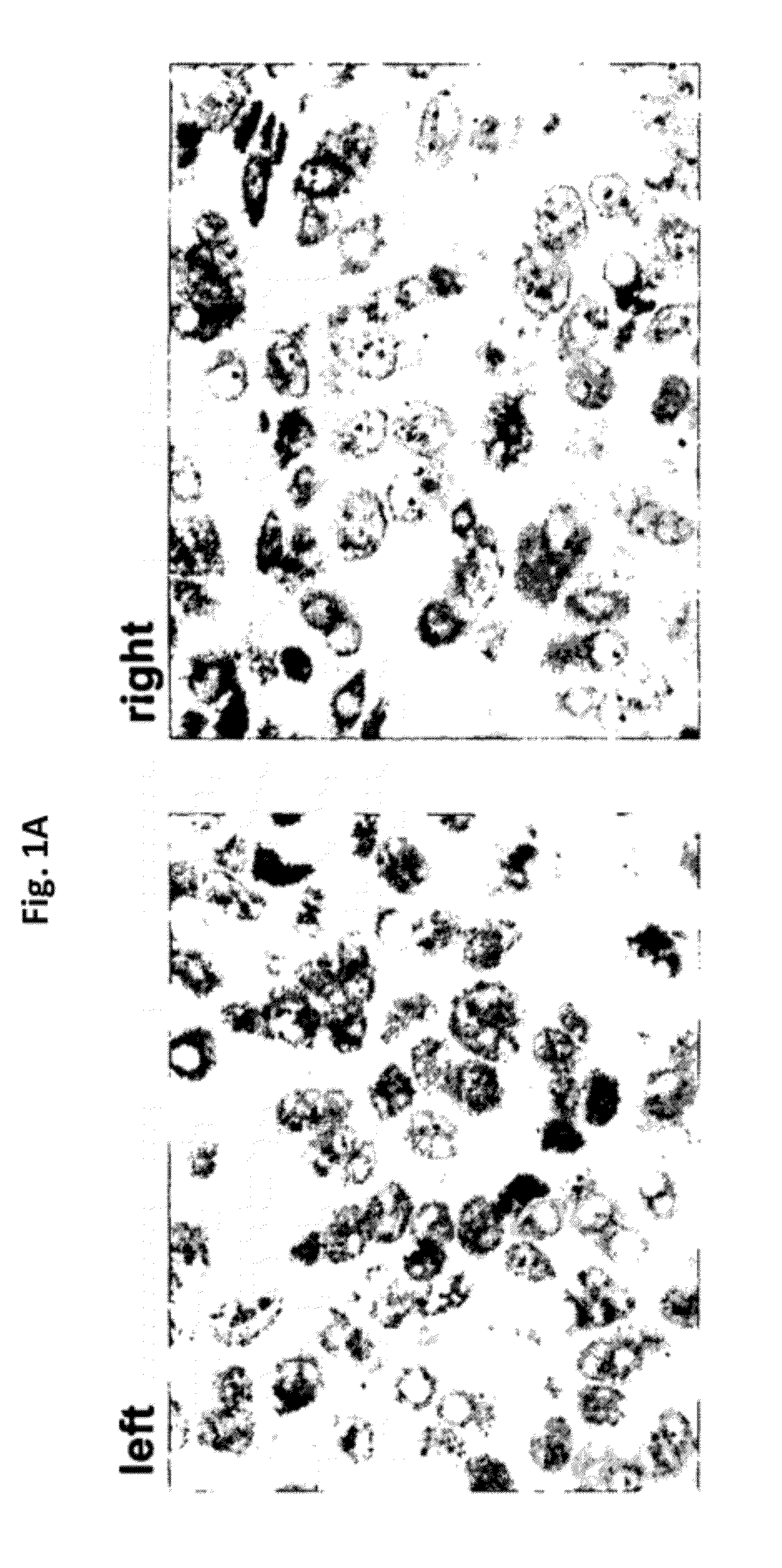
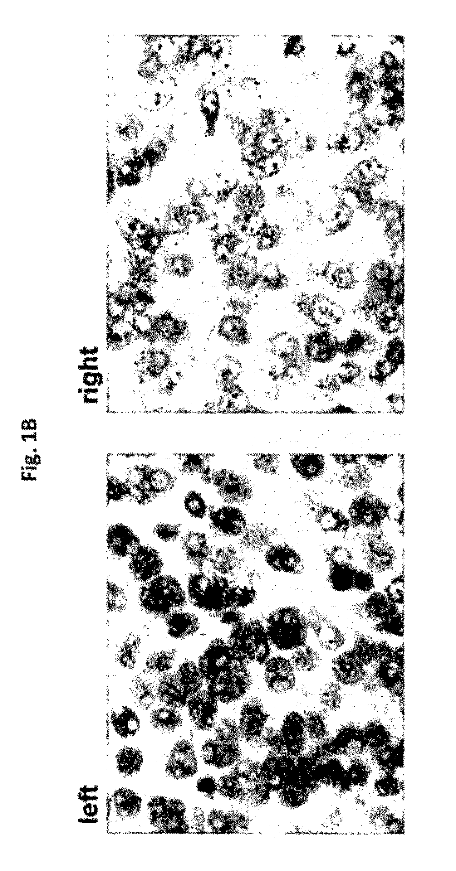
[0175]With reference to FIGS. 1A, 1B, and 1C, in situ hybridization results, using DNP-labeled oligonucleotide probes at various concentrations, for HER-2 genomic gene sequences in Xenografts derived from Calu3 cells (FIG. 1A), ZR-75-1 cells (FIG. 1B), and MCF7 cells (FIG. 1C) are presented. The various cell lines show differing gene amplification (Calu3—no gene amplification; ZR-75-1 cells—low gene amplification; MCF7 cells—high gene amplification). As demonstrated, at a hybridization time of 2 hours, the set of oligonucleotide probes according to the present disclosure resulted in similar signal intensity for all cell lines, even at the lower probe concentration (4 μg / ml vs. 10 μg / ml) when compared to the Ventana INFORM HER2 DNA Probe™ (Ventana Medical Systems Inc., USA).
example 2
[0176]With reference to FIGS. 2A, 2B, and 2C, in situ hybridization results, using DNP-labeled oligonucleotide probes, for HER-2 genomic gene sequences in Xenografts derived from ZR-75-1 cells are presented. These results were obtained following differing lengths of hybridization with the oligonucleotide probes and using enzyme metallographic detection. As demonstrated in these Figures, using the set of oligonucleotide probes according to the present disclosure, a more intense staining signal was observed after 32 minutes or after 1 hour of hybridization, as compared to the Ventana INFORM HER2DNA Probe™, at a lower concentration (4 μg / ml vs. 10 μg / ml). While the set of oligonucleotide probes according to the present disclosure resulted in optimal staining after a hybridization time of only 32 minutes, optimal staining using the INFORM™ HER2DNA Probe was achieved only after 2 hours, and suboptimal staining was achieved after 1 hour (see FIG. 2B).
example 3
[0177]With reference to FIGS. 3A and 3B, when using metallographic detection, the clustering of oligonucleotide probe subsets at both ends of the target locus (leaving the middle part of the target locus unlabeled) gives a more intense staining than an even distribution of oligonucleotide probe subsets over the entire target locus (see FIGS. 3A and 3B, Pool 2 vs. Pool 4). It is noted that the total number of labels, however, is equal in both pools. This illustrates an advantage of the set of oligonucleotide probes of the present disclosure in that it provides the possibility to design optimal compositions of oligonucleotide sets (or subsets) in order to obtain optimal signal intensity while minimizing the number of necessary oligonucleotide probe sets (or subsets).
PUM
| Property | Measurement | Unit |
|---|---|---|
| Length | aaaaa | aaaaa |
| Distance | aaaaa | aaaaa |
Abstract
Description
Claims
Application Information
 - R&D
- Intellectual Property
- Life Sciences
- Materials
- Tech Scout
- Unparalleled Data Quality
- Higher Quality Content
- 60% Fewer Hallucinations
Browse by: Latest US Patents, China's latest patents, Technical Efficacy Thesaurus, Application Domain, Technology Topic, Popular Technical Reports.
© 2025 PatSnap. All rights reserved.Legal|Privacy policy|Modern Slavery Act Transparency Statement|Sitemap|About US| Contact US: help@patsnap.com