Diagnosis and treatment of cancers with microRNA located in or near cancer associated chromosomal features

a cancer and chromosomal feature technology, applied in the field of diagnosis of cancers, can solve the problems of large amplification, deletion or rearrangement near the integration site, abnormally high gaps or breaks, and researchers are unable to develop treatments or diagnostic tests which cover more than a few types of cancer

Inactive Publication Date: 2006-05-18
THOMAS JEFFERSON UNIV
View PDF4 Cites 384 Cited by
  • Summary
  • Abstract
  • Description
  • Claims
  • Application Information

AI Technical Summary

Benefits of technology

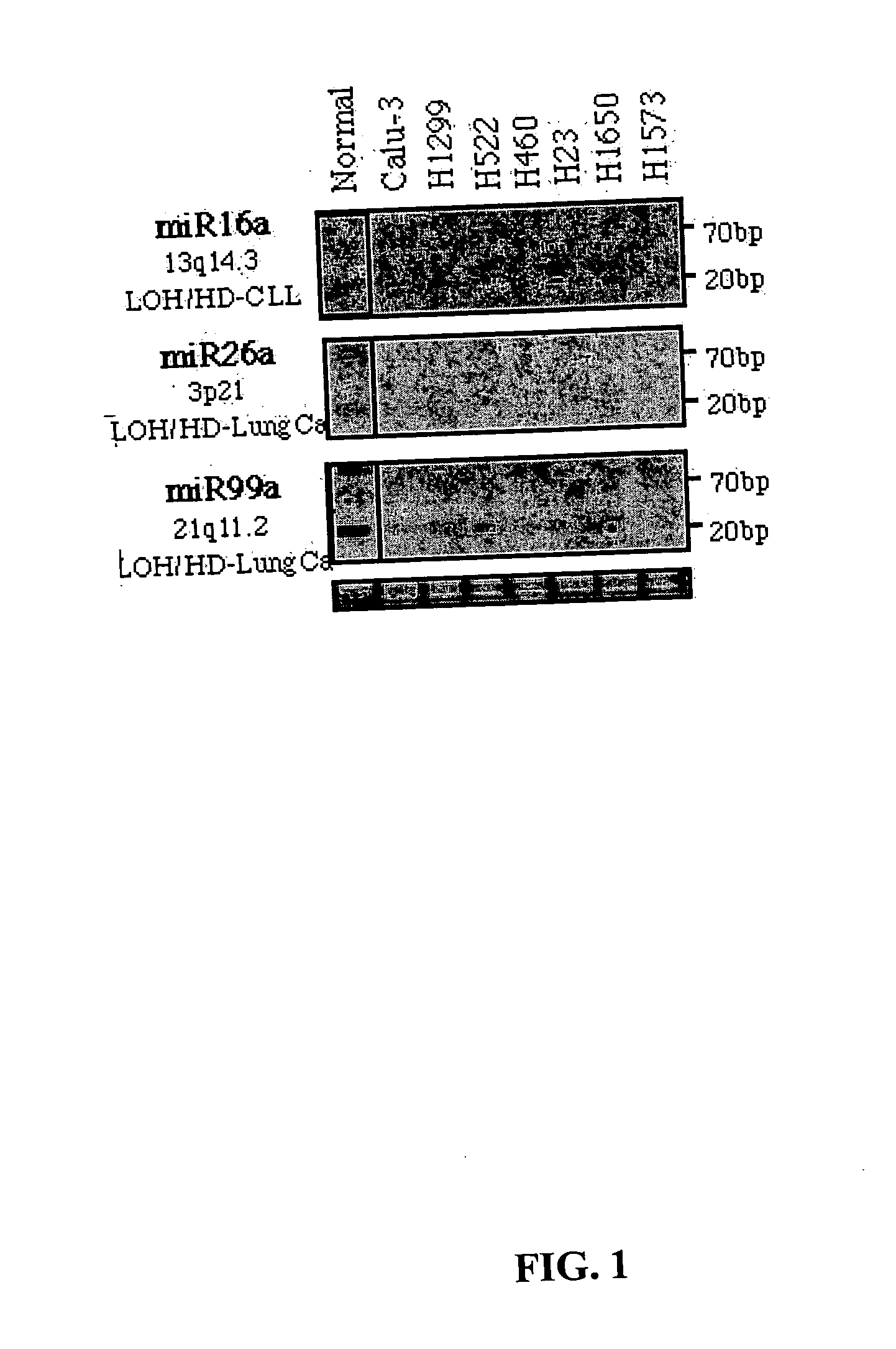
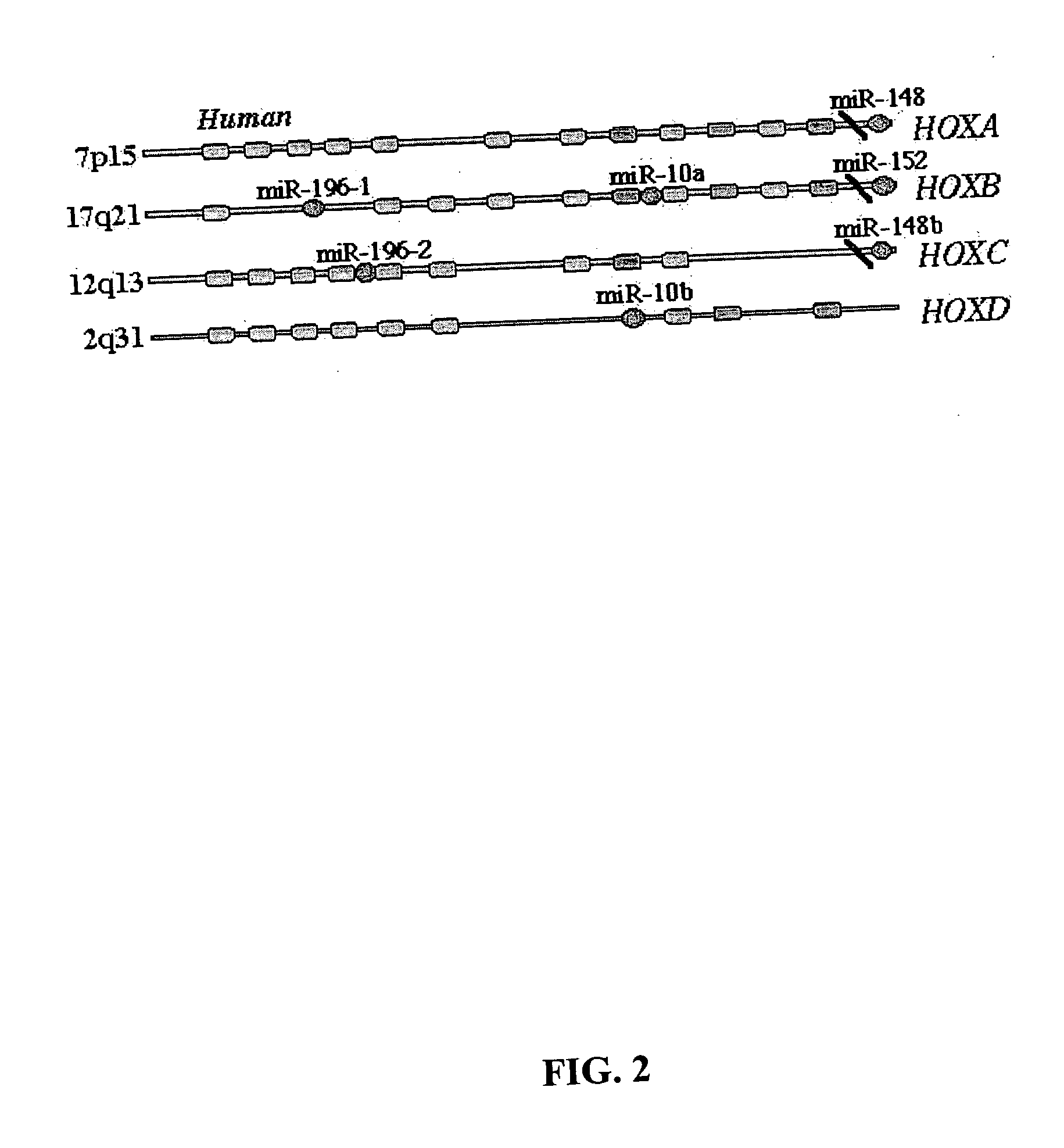
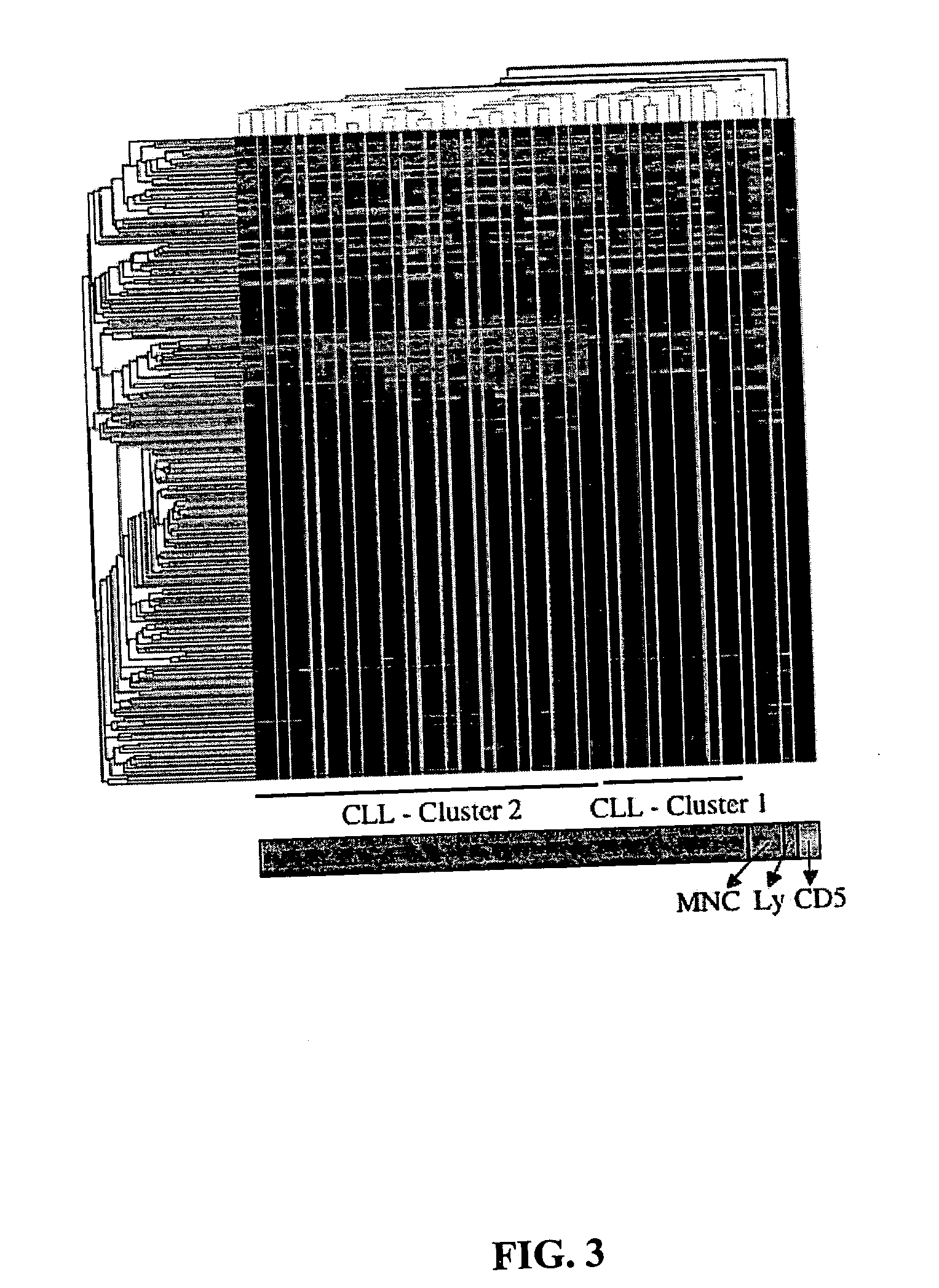
[0015] It has now been discovered that miR genes are commonly associated with chromosomal features involved in the etiology of different cancers. The perturbations in the genomic structure or chromosomal architecture of a cell caused by a cancer-associated chromosomal feature can affect the expression of the miR gene(s) located in close proximity to that chromosomal feature. Evaluation of miR gene expression can therefore be used to indicate the presence of a cancer-causing chromosomal lesion in a subject. As the change in miR gene expression level caused by a cancer-associated chromosomal feature may also contribute to cancerigenesis, a given cancer can be treated by restoring the level of miR gene expression to normal.

Problems solved by technology

Researchers therefore have been unable to develop treatments or diagnostic tests which cover more than a few types of cancer.
Chromosomal fragile sites are regions of genomic DNA which show an abnormally high occurrence of gaps or breaks when DNA synthesis is perturbed during metaphase.
HPV integration into a host cell genome can cause large amplification, deletions or rearrangements near the integration site.
However, the distribution of miR genes throughout the genome, and the relationship of the miR genes to the diverse chromosomal features discussed herein, has not been systematically studied.

Method used


Image

  • Diagnosis and treatment of cancers with microRNA located in or near cancer associated chromosomal features
  • Diagnosis and treatment of cancers with microRNA located in or near cancer associated chromosomal features
  • Diagnosis and treatment of cancers with microRNA located in or near cancer associated chromosomal features

Examples


example 1

miR Genes are Non-Randomly Distributed in the Human Genome

[0175] One hundred eighty-six human genes representing known or predicted miR genes were mapped, based on mouse homology or computational methods, as described above in the General Methods. The results are presented in Table 2. The names were as in the miRNA Registry; for new miR genes, sequential names were assigned. miR 213 from Sanger database is different from miR 213 described in Lim et al. (2003, Science 299:1540). MiR genes in clusters are separated by a forward slash “ / ”. The approximate location in Mb of each clone is presented in the last column.

TABLE 2miR Database: Chromosome Location and ClusteringChromosomeLoc (Mb)NamelocationGenes in Cluster(built 33)let-7a-109q22.2let-7a-1 / let-7f-1 / let-7d90.2-.3let-7a-211q24.1miR-125b-1 / let-7a-2 / miR-100  121.9-122.15let-7a-322q13.3let-7a-3 / let-7b44.7-.8let-7b22q13.3let-7a-3 / let-7b44.7-.8let-7c21q11.2miR-99a / let-7c / miR-125b-216.7-.9let-7d09q22.2let-7a-1 / let-7f-1 / let7d90.2-.3l...

example 2

miR Genes are Located in or Near Fragile Sites

[0179] Thirty-five of 186 miRs (19%) were found in (13 miR genes), or within 3 Mb (22 miR genes) of cloned fragile sites (FRA). A set of 39 fragile sites with available cloning information was used in the analysis. Data were available for the exact dimension (mean 2.69 Mb) and position of ten of these cloned fragile sites (see General Methods above). The relative incidence of miR genes inside fragile sites occurred at a rate 9.12 times higher than in non-fragile sites (p<0.001, using mixed effect Poisson regression models; see Tables 3 and 4). The same very high statistical significance was also found when only the 13 miRs located exactly inside a FRA or exactly in the vicinity of the “anchoring” marker mapped for a FRA were considered (IRR=3.21, p<0.001). Among the four most active common fragile sites (FRA3B, FRA16D, FRA6E, and FRA7H), the data demonstrate seven miRs in (miR-29a and miR-29b) or close (miR-96, miR-182s, miR-182as, miR-...

example 3

miR Genes are Located in or Near Human Papilloma Virus (HPV) Integration Sites

[0181] Because common fragile sites are preferential targets for HPV16 integration in cervical tumors, and infection with HPV16 or HPV18 is the major risk factor for developing cervical cancer, the association between miR gene locations and HPV16 integration sites in cervical tumors was analyzed. The data indicate that thirteen miR genes (7%) are located within 2.5 Mb of seven of seventeen (45%) cloned integration sites. The relative incidence of miRs at HPV16 integration sites occurred at a rate 3.22 times higher than in the rest of the genome (p<0.002) (Tables 4 and 5). In one cluster of integration sites at chromosome 17q23, where three HPV16 integration sites are spread over roughly 4 Mb of genomic sequence, four miR genes (miR-21, miR-301, miR-142s and miR-142as) were found.

TABLE 5Analyzed FRA Sites, Cancer Correlation and HPV Integration SitesDistanceLocationmiR-HPV16SymbolChromosomeCancer correla...


PUM

PropertyMeasurementUnit
weightaaaaaaaaaa
volumeaaaaaaaaaa
number-average molecular weightaaaaaaaaaa

Abstract

MicroRNA genes are highly associated with chromosomal features involved in the etiology of different cancers. The perturbations in the genomic structure or chromosomal architecture of a cell caused by these cancer-associated chromosomal features can affect the expression of the miR gene(s) located in close proximity to that chromosomal feature. Evaluation of miR gene expression can therefore be used to indicate the presence of a cancer-causing chromosomal lesion in a subject. As the change in miR gene expression level caused by a cancer-associated chromosomal feature may also contribute to cancerigenesis, a given cancer can be treated by restoring the level of miR gene expression to normal. microRNA expression profiling can be used to diagnose cancer and predict whether a particular cancer is associated with an adverse prognosis. The identification of specific mutations associated with genomic regions that harbor miR genes in CLL patients provides a means for diagnosing CLL and possibly other cancers.

Description

RELATED APPLICATIONS [0001] This application is a continuation-in-part of International Application No. PCT / US2005 / 004865, filed Feb. 9, 2005, which claims the benefit of U.S. Provisional Application No. 60 / 543,119, filed Feb. 9, 2004, U.S. Provisional Application No. 60 / 542,929, filed Feb. 9, 2004, U.S. Provisional Application No. 60 / 542,963, filed Feb. 9, 2004, U.S. Provisional Application No. 60 / 542,940, filed Feb. 9, 2004, U.S. Provisional Application No. 60 / 580,959, filed Jun. 18, 2004, and U.S. Provisional Application No. 60 / 580,797, filed Jun. 18, 2004. The entire teachings of the above applications are incorporated herein by reference.GOVERNMENT SUPPORT [0002] The invention described herein was supported in part by grant nos. P01CA76259, P01CA81534, and P30CA56036 from the National Cancer Institute. The U.S. government has certain rights in this inventionFIELD OF THE INVENTION [0003] The invention relates to the diagnosis of cancers, or the screening of individuals for the p...

Claims


Application Information

Patent Timeline
Patent Type & Authority Applications(United States)
IPC IPC(8): C12Q1/68
CPCC12Q1/6886C12Q2600/106C12Q2600/158C12Q2600/178A61K31/7088A61K31/713A61K45/06C12N15/113A61P35/00C12N15/1135C12N2310/11C12N2310/31C12N2310/32C12N2310/33C12N7/00C12N2310/141C12N2710/16143
Inventor CROCE, CARLOLIU, CHANG-GONGCALIN, GEORGESEVIGNANI, CINZIA
Owner THOMAS JEFFERSON UNIV
Why Eureka
Try Eureka