
Minimally invasive apparatus and method for treating the retina

Inactive Publication Date: 2017-04-20
MAZZOCCHI RUDY A +1
View PDF0 Cites 5 Cited by
  • Summary
  • Abstract
  • Description
  • Claims
  • Application Information

AI Technical Summary

Benefits of technology

The invention is a method and device for treating retinal detachments by placing a permeable structure on the back of the eye to hold the retina in place. This can help to restore some vision and prevent further damage to the retina. The device is inserted through an incision in the eye and fixed in place. The method can be temporary or permanent, depending on the needs of the patient.

Problems solved by technology

Such devices are commonly used in the cardiology and peripheral vasculature fields to open vessels or to capture particles in circulating fluid but are not known for use within delicate organs such as the eye.

Method used


Image

  • Minimally invasive apparatus and method for treating the retina
  • Minimally invasive apparatus and method for treating the retina
  • Minimally invasive apparatus and method for treating the retina

Examples


Embodiment Construction

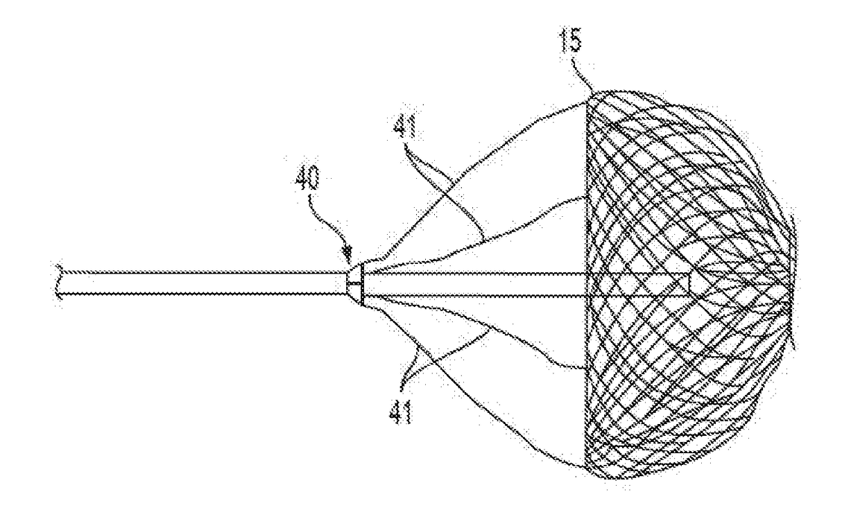
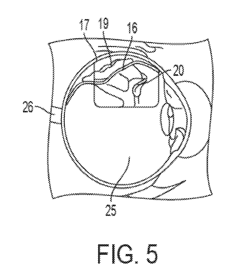
[0032]A retinal detachment is a separation of the retina from its attachments to its underlying supporting choroids tissue within the eye. Most retinal detachments are a result of an opening in the retinal wall due to a break, hole, or tear. The opening in the retina may allow the vitreous humor fluid to get in between the retina and the choroid. The fluid can force the retina away from the choroid causing blind spots where the retina has separated. Where the retina is weak, it will tear. As more of the vitreous collects behind the retina, the extent of the retinal detachment can progress and possibly involve the entire retina, leading to a total retinal detachment. A representative tear 20 is shown in FIG. 5 in which the retina 16 has separated from the underlying supporting choroid tissue 17. The tear 20 allows fluid 19 from the posterior chamber 25 to penetrate behind the retina 16 thereby separating the retina 16 from the underlying optic nerve 26. As fluid 19 builds up, the det...


PUM

PropertyMeasurementUnit
intra ocular pressureaaaaaaaaaa
diameteraaaaaaaaaa
superelasticaaaaaaaaaa

Abstract

The present invention comprises an implant for placing inside the eye such that the implant comes into contact with the interior tissue of the eye such that it conforms to the inner globe geometry of the eye. Implants of the present invention may also be used to alter the focal length of the eye thereby providing a treatment method for the correction of myopia and hyperopia. The device may consist of several possible configurations, an open mesh structure, a solid metal ring, a solid polymer shape, a mesh polymer shape or combination of these. The shape may be a curve, a sphere, a ring or a combination thereof that are specifically shaped to approximate a desired portion of the interior globe of the eye in order to treat myopia or hyperopia

Description

[0001]This application is a continuation in part of U.S. patent application Ser. No. 13 / 795,312 filed on Oct. 3, 2012, now U.S. Pat. No. 9,532,902 issued on Jan. 3, 2017 which is a national state entry from PCT / US2011 / 025686 filed on Feb. 22, 2011, which claims priority to provisional patent application No. 61 / 306,644 filed on Feb. 22, 2010, the contents of which are expressly incorporated herein by reference. All references cied herein are expressly incorporated by reference.[0002]The retina is the interior layer of the posterior eyeball. The retina receives images as they enter through the lens, is continuous with the optic nerve, and consists of several layers, one of which contains the rod cells and cone cells that are sensitive to light.[0003]A retinal detachment is a separation of the retina from its attachments to the underlying supporting choroid tissue within the eye. Most retinal detachments are a result of an opening that originates as a retinal break, hole, or tear. This...

Claims


Application Information

Patent Timeline
Patent Type & Authority Applications(United States)
IPC IPC(8): A61F9/007A61F2/00
CPCA61F9/00727A61F2/0063A61F2210/0004A61F2002/0068A61F2250/0091A61F2002/0072A61F2210/0014A61F2/14
Inventor MAZZOCCHI, RUDY ACALHOUN, MICHAEL W.
Owner MAZZOCCHI RUDY A
Learn More