Method and system to digitize pathology specimens in a stepwise fashion for review

a pathology and stepwise technology, applied in the field of digital light microscopy, can solve the problems of slow adoption of digital or “virtual” microscopy, slow image viewing, cumbersome and slow,

Active Publication Date: 2014-01-30
THE JOHN HOPKINS UNIV SCHOOL OF MEDICINE
View PDF14 Cites 6 Cited by
  • Summary
  • Abstract
  • Description
  • Claims
  • Application Information

AI Technical Summary

Benefits of technology

[0008]A method for reviewing digitized images of pathology specimens according to an embodiment of the current application includes receiving a whole-slide digital image of a pathology specimen, segmenting the whole-slide digital image into a tissue-particle image within the whole-slide digital image, and representing the tissue-particle image as a plurality of image tiles such that each of the plurality of image tiles can be displayed sequentially on an image display system to permit a substantially complete, tile-by-tile review of the tissue-particle image in a predefined order.

Problems solved by technology

Adoption of digital or “virtual” microscopy has been slow.
While there have been improvements in the speed and quality of scanning hardware, image viewers and viewing techniques have remained stagnant.
Compared to the relative ease of operating a microscope, navigating digital slides is unintuitive, cumbersome and slow.
This results in lost productivity and user frustration.

Method used


Image

  • Method and system to digitize pathology specimens in a stepwise fashion for review
  • Method and system to digitize pathology specimens in a stepwise fashion for review
  • Method and system to digitize pathology specimens in a stepwise fashion for review

Examples


Embodiment Construction

[0014]Some embodiments of the current invention are discussed in detail below. In describing embodiments, specific terminology is employed for the sake of clarity. However, the invention is not intended to be limited to the specific terminology so selected. A person skilled in the relevant art will recognize that other equivalent components can be employed and other methods developed without departing from the broad concepts of the current invention. All references cited anywhere in this specification, including the Background and Detailed Description sections, are incorporated by reference as if each had been individually incorporated.

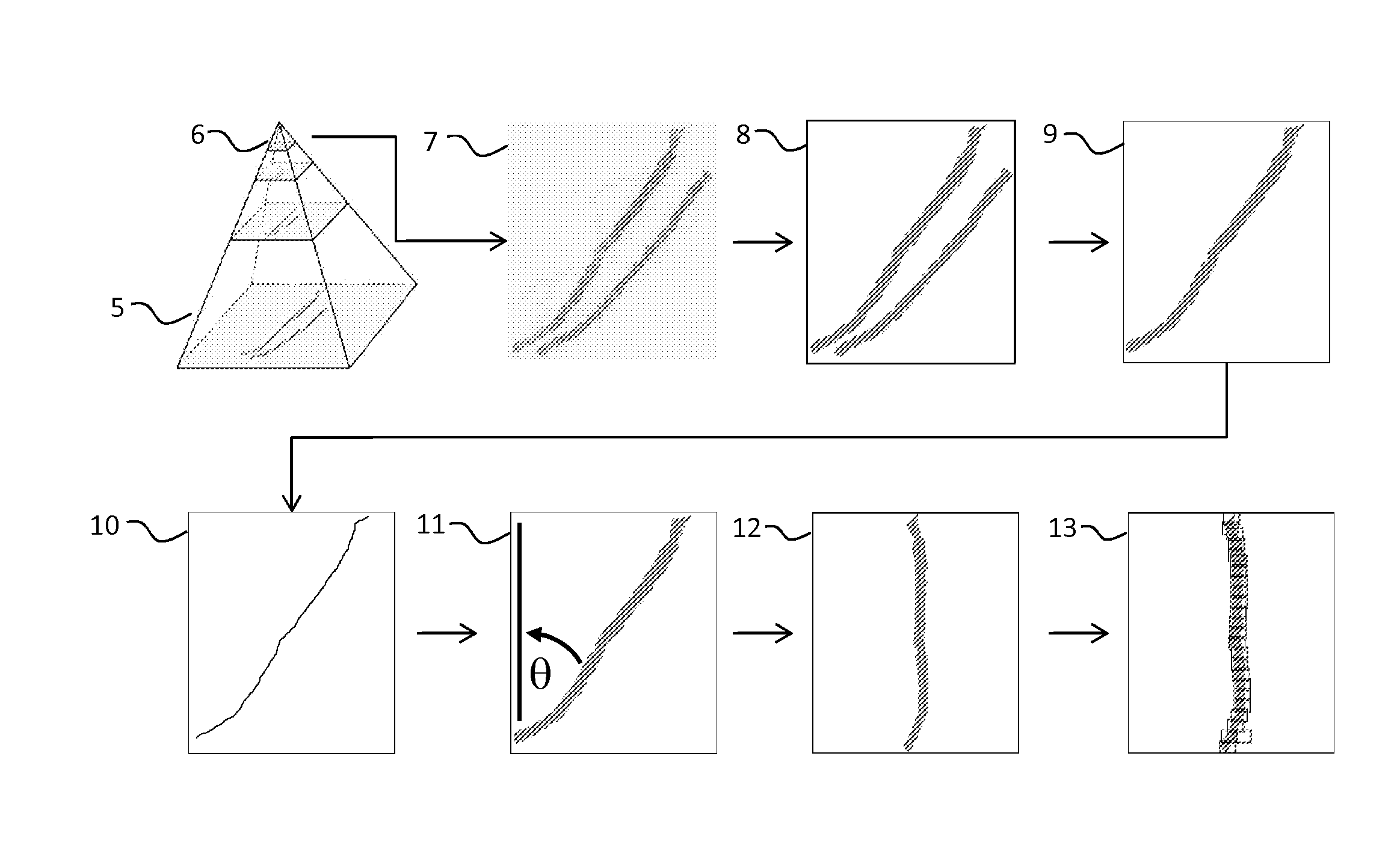
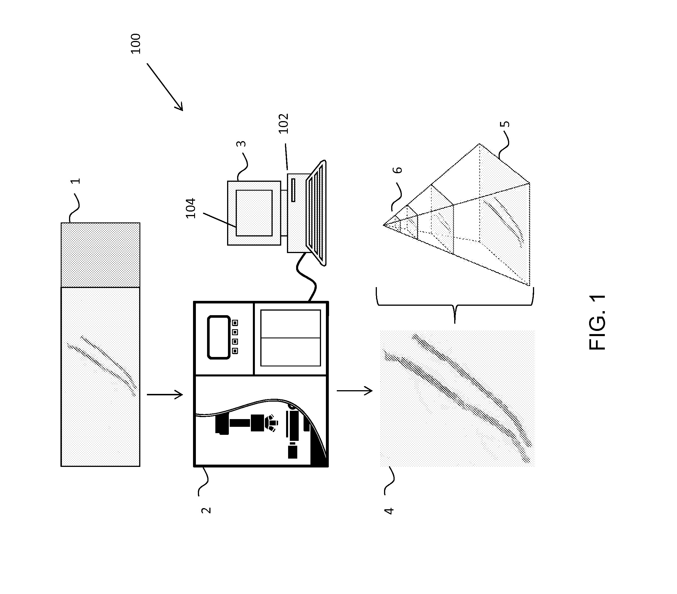
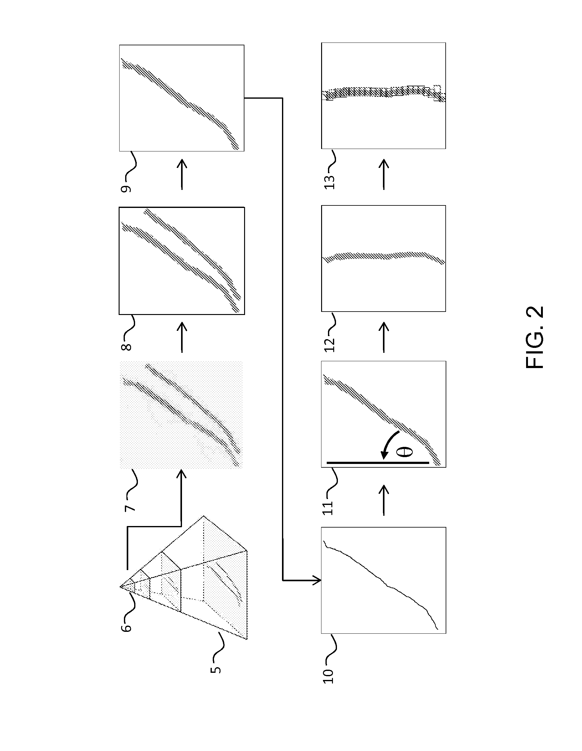
[0015]If the promise of digital or “virtual” microscopy is to be fully realized, then we must have tools that are as good as, or better than current methods of slide evaluation. Currently, microscopists interact directly with a glass slide, using their fingers or an X-Y stage to move the slide while focusing in the Z-axis. In practice, rather than vie...


PUM


Abstract

A system for reviewing digitized images of pathology specimens includes an image processing system and an image display system adapted to communicate with the image processing system. The image processing system is configured to receive a whole-slide digital image of a pathology specimen, segment the whole-slide digital image into a tissue-particle image within the whole-slide digital image, and represent the tissue-particle image as a plurality of image tiles such that each of the plurality of image tiles can be displayed sequentially on the image display system to permit a substantially complete, tile-by-tile review of the tissue-particle image in a predefined order.

Description

CROSS-REFERENCE OF RELATED APPLICATION[0001]This application claims priority to U.S. Provisional Application No. 61 / 443,022 filed Feb. 15, 2011, the entire contents of which are hereby incorporated by reference.BACKGROUND[0002]1. Field of Invention[0003]The field of the currently claimed embodiments of this invention relates to digital light microscopy, and more particularly to systems and methods for reviewing digitized images of pathology specimens.[0004]2. Discussion of Related Art[0005]The standard method of examining tissue sections under a microscope requires a number of preliminary steps. Specimens are first acquired from human or animal tissues and then are typically fixed in formalin and embedded in paraffin. Thin tissue sections are cut from the paraffin blocks and mounted on glass slides. The tissue sections are stained with chemical and / or antibody-based staining reagents that highlight structures of interest. To review the slide digitally, it is then digitized using a s...

Claims


Application Information

Patent Timeline
Patent Type & Authority Applications(United States)
IPC IPC(8): G06T7/00
CPCG06T7/0012G06T2207/10056G06T2207/20016G06T2207/30024G06T3/40G06V20/693
Inventor HALUSHKA, MARC KENNETHCORNISH, TOBY CHARLES
Owner THE JOHN HOPKINS UNIV SCHOOL OF MEDICINE
Why Eureka
Try Eureka