Apparatus, system and methods for proper transesophageal echocardiography probe positioning by using camera for ultrasound imaging

a technology of ultrasound imaging and esophageal echocardiography, which is applied in the field of transesophageal echocardiography endoscopic camera assist devices, can solve the problems of increasing tee related complications, low reported complication rates, and increasing so as to reduce or reduce the risk of esophageal and pharyngeal complications, the effect of minimizing or reducing the risk o

Active Publication Date: 2016-06-02
VISURA TECH LLC
View PDF12 Cites 15 Cited by
  • Summary
  • Abstract
  • Description
  • Claims
  • Application Information

AI Technical Summary

Benefits of technology

[0009]The present disclosure solves the disadvantages of, and improves on, the prior art systems as described herein. The present disclosure pertains to a device, system and methods that comprise adding a removable or detachable camera to a TEE probe, for example, by using a carrier comprised of a proximal component, a mid-section lumen and a distal component, and in which the distal component is configured to removeably attach to the distal end of the TEE probe. The distal component houses a camera and light source or LED and associated wiring back to the proximal component through the mid-section lumen for connection to a video monitor to allow the practitioner to view the placement of the TEE probe in the patient, thereby minimizing or reducing the risk of esophageal and pharyngeal complications during the positioning of the TEE probe into the patient's esophagus. The reduction or minimization of complications occurs by allowing the cardiologist direct visualization of pharyngeal and esophageal structures during intubation. This device and procedure eliminates any “blind procedure”, whereby direct visualization of the pharyngeal structures and esophagus present a possible solution to these mechanical complications. Once the TEE probe is properly located, the carrier can be removed from the TEE probe and extracted from the patient.
[0012]A device, system and methods that provide the above-noted advantages is the TEECAD, a Transesophageal Echocardiography Endoscopic Camera Assist Device, which offers a way to allow the cardiologist direct visualization for TEE intubation, without having to develop a completely new TEE probe or revamp existing echocardiography machines to accommodate the technology. As detailed herein, the TEECAD device comprises a carrier with a camera and light source device and system that is configured to be attached to, and when necessary, removed from, an existing probe. The carrier or attachment device can be further configured with wiring to transmit information to a video monitor, which may be separate from the echocardiography machine. The device can also be used with an eyepiece for the cardiologists to look into during the placement of the probe. An advantage to the TEECAD device is the minimal added width and / or girth on the TEE probe, and the device is configured to be detachable from the probe and easily retractable after successful intubation of the esophagus.
[0014]This configuration allows the practitioner to view the placement of the TEE probe in the patient as the TEECAD device would be configured to feed video of the probe placement (in the esophagus) on a separate portable monitor, which can be placed near the device dependent on the operator's preference. The distal component would be configured to be held in place on the back end of the TEE probe and could be removed with the correct amount of axial tension, once the TEE probe was successfully located in the esophagus. Once detached from the TEE probe, the TEECAD device could be retracted and removed from the patient's esophagus.
[0015]In the preferred embodiment, the distal component would be configured to house the small video camera, lens, and light source, such as an LED. The internal wiring will comprise of a video feed wire, a power source for the camera and LED. A control handle at the end of the device would provide any needed controls for the video feed and for the LED. The camera can also be wireless, and the light source and camera may contain an internal power supply, to reduce the need for some or all of the wires.

Problems solved by technology

Reported complication rates are generally low, with the most catastrophic potential complication being perforation of the esophagus or damage to pharyngeal structures.
While the reported complication rates appear low, these rates are likely to change as the risk profile of patients undergoing TEE evolves, especially with the advancing age of patients undergoing TEE, which will almost certainly lead to an increase in TEE related complications, such as esophageal and pharyngeal perforation, and accidental tracheal intubation.

Method used


Image

  • Apparatus, system and methods for proper transesophageal echocardiography probe positioning by using camera for ultrasound imaging
  • Apparatus, system and methods for proper transesophageal echocardiography probe positioning by using camera for ultrasound imaging
  • Apparatus, system and methods for proper transesophageal echocardiography probe positioning by using camera for ultrasound imaging

Examples


Embodiment Construction

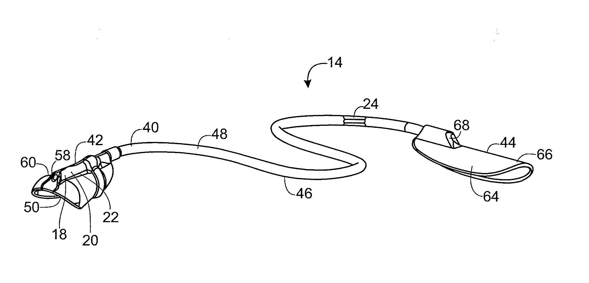
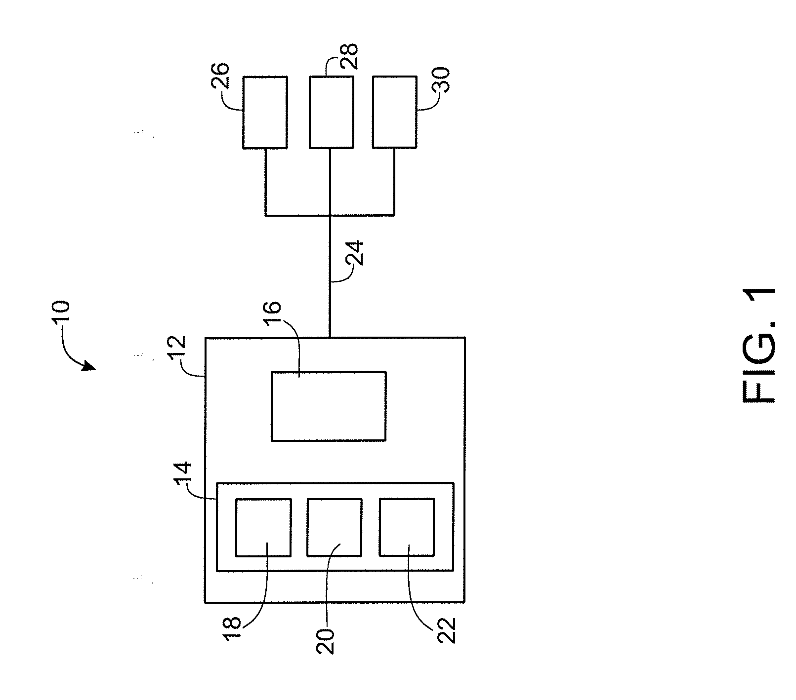
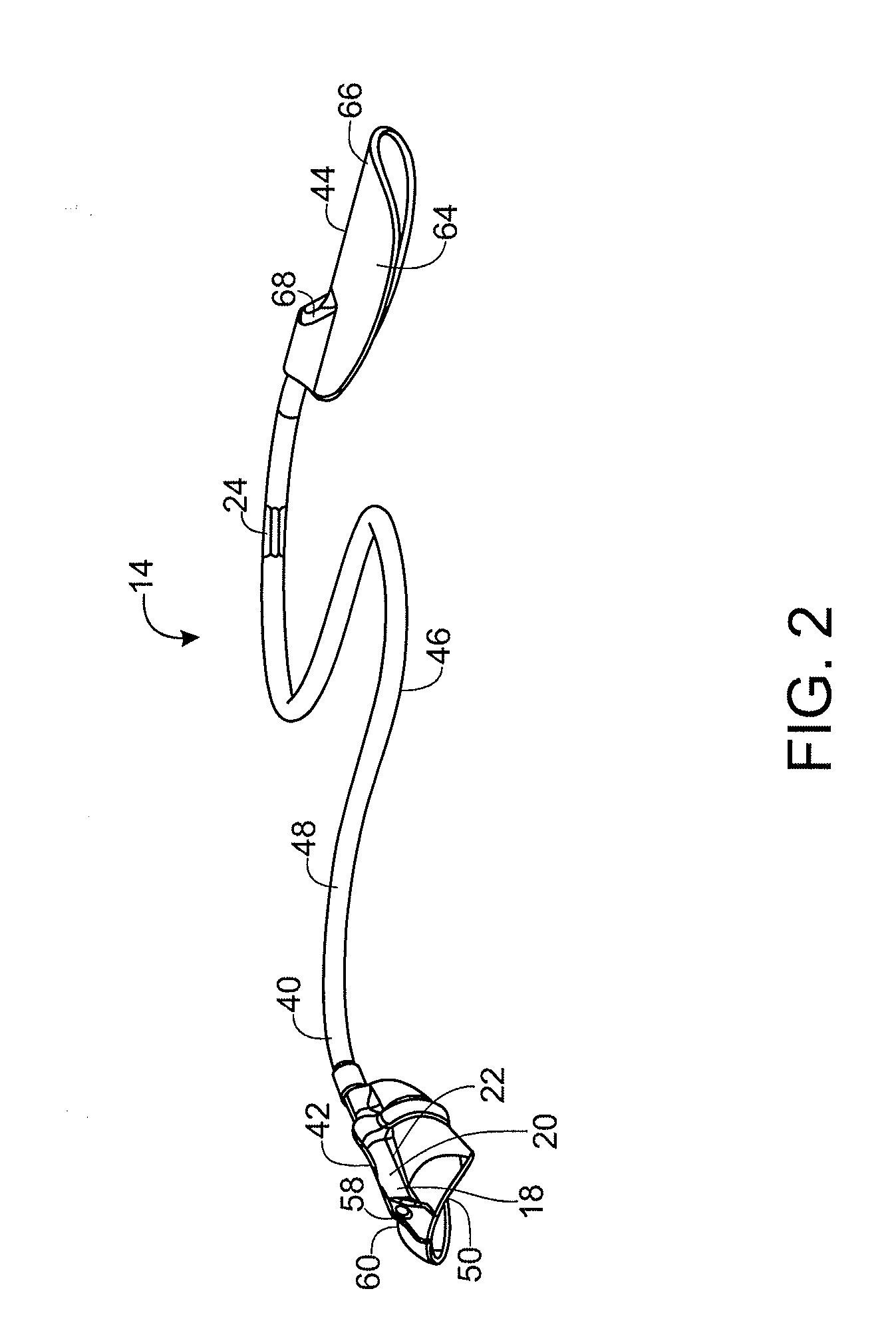
[0024]The present disclosure is an apparatus, system and related methods pertaining to an attachable and detachable TEECAD or Transesophageal Echocardiography Endoscopic Camera Assist Device. The TEECAD device increases the functionality of currently existing probes by transmitting video information to a video monitor during the placement of the probe. The video information can be displayed on a video monitor, which may or may not be separate from the echocardiography machine. In particular, the display monitor used during the transesophageal echocardiography procedure, allows the practitioner or cardiologist to see or visualize the path of the TEE probe as it is inserted into the esophagus and moved into position for the TEE procedure. The TEECAD device has minimal circumference or girth and would not create an issue during intubation. Further, the TEACAD device is configured to be completely detachable, and easily retractable after a successful intubation of the esophagus, while l...


PUM


Abstract

An apparatus, system and methods that comprise adding a removable or detachable carrier containing a camera and illumination to a TEE probe, thereby allowing the user to view the placement of the probe and minimize or reduce the risk of esophageal and pharyngeal complications during the positioning of the TEE probe into the patient's esophagus. The reduction or minimization of complications occurs by allowing the cardiologist direct visualization of pharyngeal and esophageal structures during intubation. This device and procedure eliminate any “blind procedure”, whereby direct visualization of the pharyngeal structures and esophagus present a possible solution to these mechanical complications. Once the TEE probe has been properly positioned in the esophagus, the carrier is removed from the esophagus. The removable or detachable camera of the present disclosure can be used in other industries.

Description

FIELD OF THE INVENTION[0001]The present disclosure relates generally to a Transesophageal Echocardiography Endoscopic Camera Assist Device (TEECAD) for adding direct visualization to Transesophageal Echocardiography intubation (TEE), without having to develop a completely new TEE probe nor without the need to revamp existing echocardiography machines. In particular, the present disclosure comprises an attachable TEECAD probe, which is configured to be attached to an existing TEE probe prior to intubation. The TEECAD probe contains a camera and a light source, which will feed into a video monitor and allow the user or practitioner to visualize the path of the TEE probe as it is inserted into the esophagus and moved into position for the procedure. The TEECAD probe would add minimal width or girth to the TEE probe, and would be completely detachable and easily retractable after successful intubation of the esophagus, while leaving the probe in the proper location.BACKGROUND OF THE INV...

Claims


Application Information

Patent Timeline
Patent Type & Authority Applications(United States)
IPC IPC(8): A61B1/05A61B1/267A61B1/06A61B1/00
CPCA61B1/053A61B1/00018A61B1/00016A61B1/06A61B1/00103A61B1/267A61B1/00045A61B1/0676A61B1/0684A61B1/2733A61B8/0883A61B8/4455A61B1/00142A61B8/12
Inventor MARMOR, DAVID B.
Owner VISURA TECH LLC
Why Eureka
Try Eureka