
Anti-par2 antibodies and uses thereof
- Summary
- Abstract
- Description
- Claims
- Application Information
AI Technical Summary
Benefits of technology
Problems solved by technology
Method used
Image

Examples
example 1
Generation of Antibodies with pH Sensitive Binding for PAR2
[0146]Generation of Recombinant Human, Rat and Cynomolgus PAR2 and PAR1 Proteins.
[0147]The human, rat and cynomolgus (Macaca fascicularis) PAR2 (Proteinase-Activated Receptor 2) constructs comprising extracellular residues 1-75 were designed with N-terminal AviTag™ (Avidity LLC) and C-terminal Flag and poly Histidine tags and cloned into the vector pDEST12.2 OriP FH (Life Technologies). The human PAR1 (Proteinase-Activated Receptor 1) construct comprising extracellular residues 1-102 was designed with C-terminal Flag and poly Histidine tags and cloned into the vector pDEST12.2 OriP FH (Life Technologies). The constructs were expressed in HEK293 cells and purified from the media using standard affinity and size exclusion chromatography purification. To generate biotinylated proteins the AviTag™ was biotinylated enzymatically according to the manufacturer's instructions.
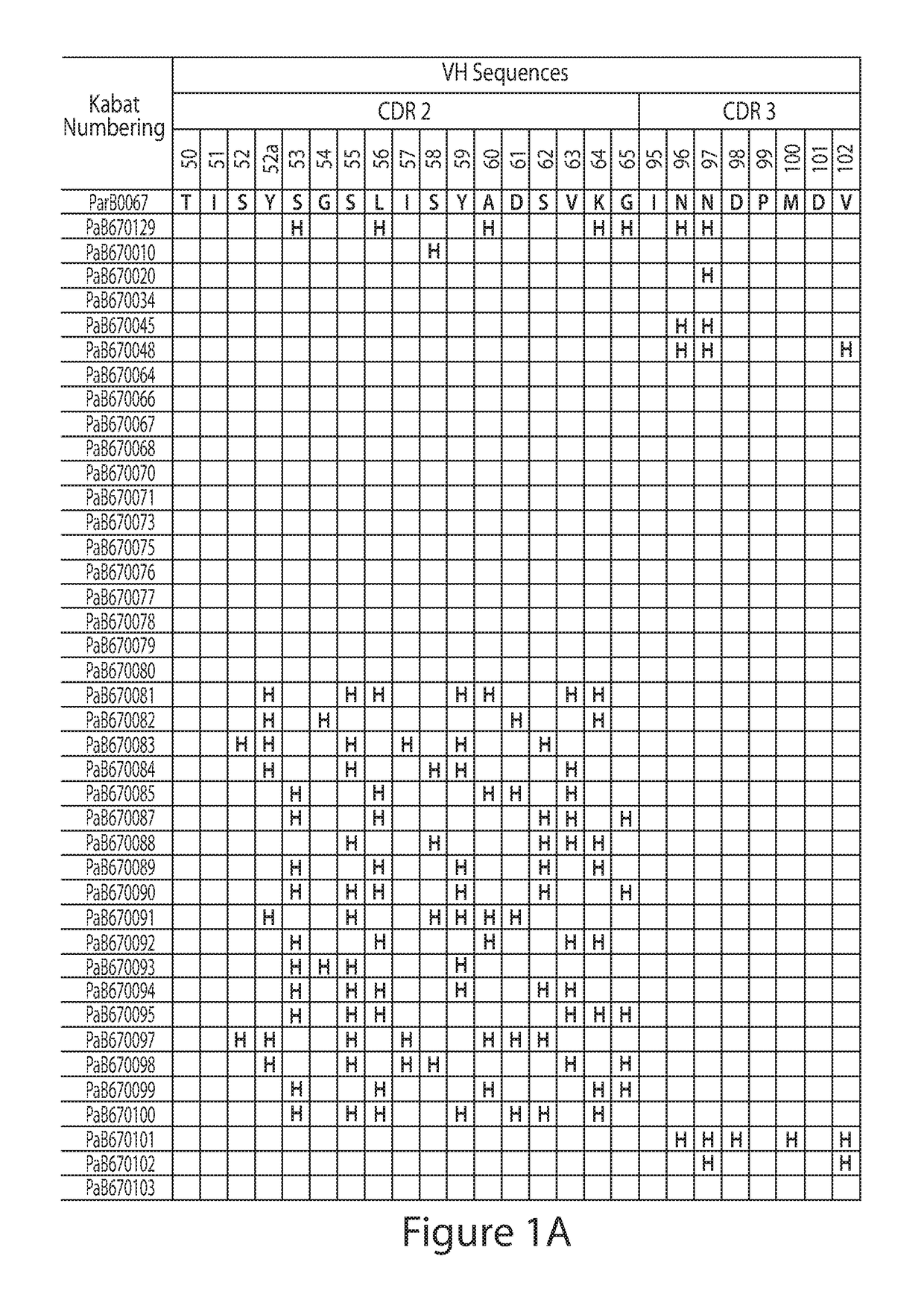
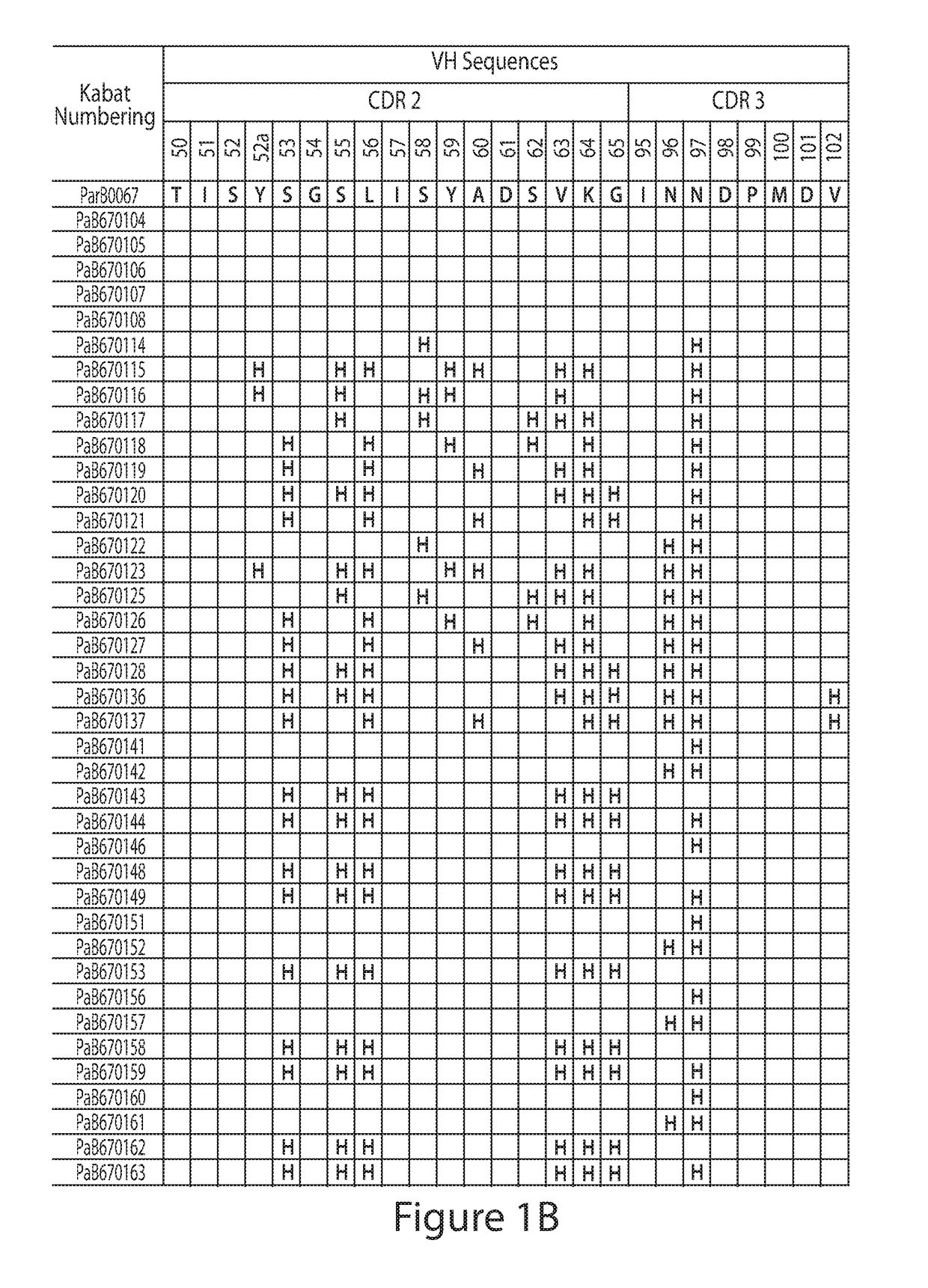
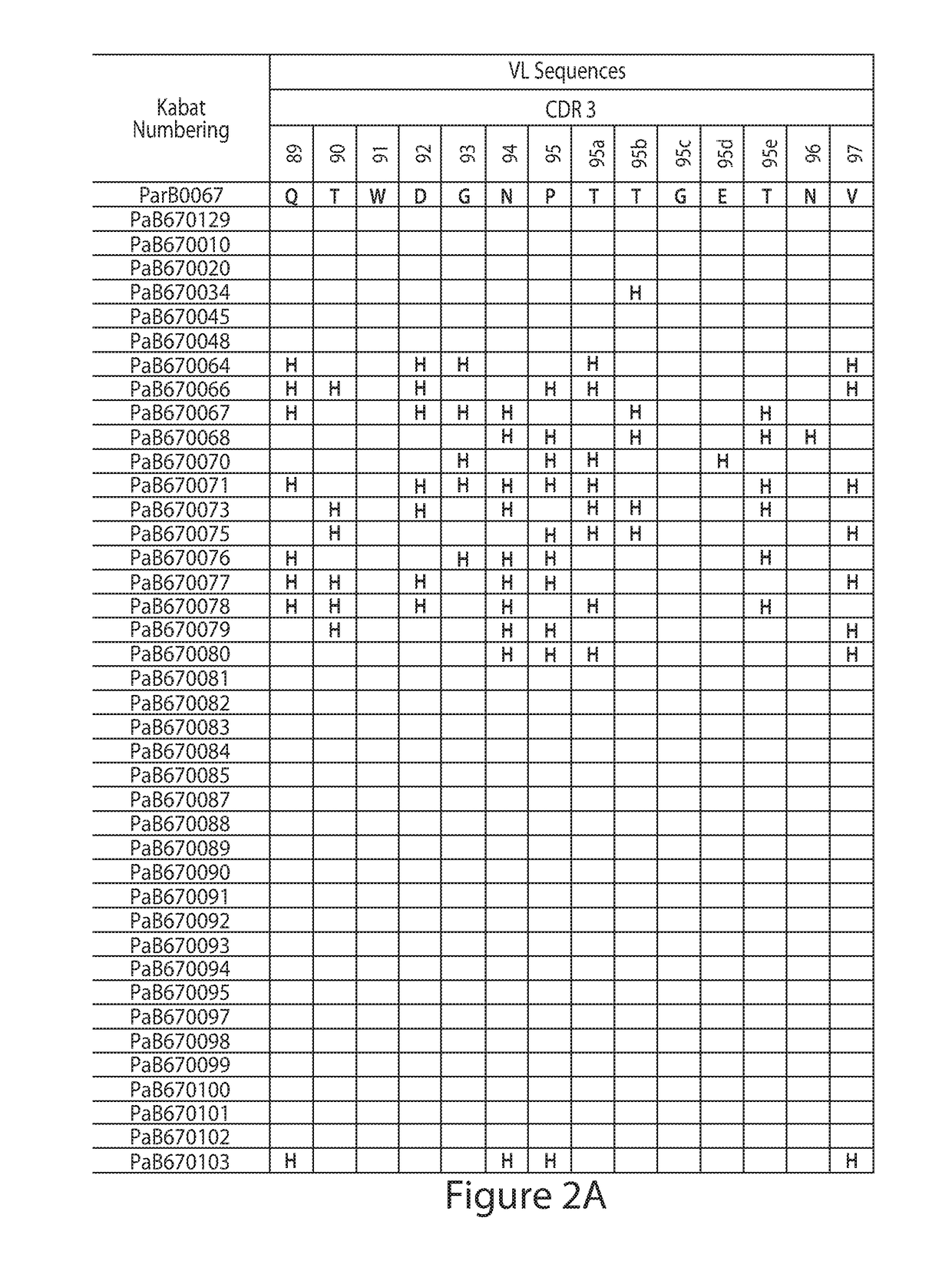
[0148]Construction of Combinatorial Histidine Scanning Li...
example 2
Cell-Based PAR2 and PAR1 Activity Assay
[0205]Human A549 cells, rat KNRK, mouse LL / 2 or cynomolgus CYNOM-K1 cells expressing endogenous PAR2, or human 1321N1-hPAR2-c18 cells overexpressing human PAR2 were seeded at 5,000 (human, cyno) or 7,000 (mouse, rat) cells per well on PDL-coated Tissue Culture Plates, (Greiner Bio-One). Cells were loaded with Fluo-Screen Quest™ Fluo-8 No Wash Calcium dye (AAT Bioquest, Inc). Cells were pre-treated with IgGs or Fabs diluted in assay buffer (HBSS, 0.1% BSA, 20 mM HEPES) for 1 h at room temperature. For mouse and cyno assays, cells were pre-treated with 0.5 nM or 10 nM thrombin, respectively, to desensitize PAR1 activity. PAR2 calcium responses to 11 nM (human), 400 nM (mouse) or 80 nM (rat, cyno) trypsin (Polymun) were measured on a Fluorescent Imaging Plate Reader (FLIPR) Tetra (Molecular Devices). To determine functional activity against human PAR1, thrombin driven calcium responses in the A549 human cell line were determined also on the FLIPR ...
example 3
Effects of an Anti-PAR2 Antibody in a Rat Model of Inflammatory Joint Pain
[0209]Intra-articular administration of Monosodium Iodoacetate (MIA) in the ipsilateral knee of Sprague Dawley rats leads to development of a robust and long-lasting hyperalgesia and allodynia associated initially with an inflammatory response. The development of these signs in this animal model are believed to be clinically relevant; reflecting the symptoms displayed by patients presenting with chronic inflammatory pain associated with underlying conditions such as osteoarthritis (OA) or rheumatoid arthritis (Bove et al., 2003; Fernihough et al., 2004; Kalbhen 1987). It has previously been demonstrated that (using weight-bearing as an end-point) the time course of MIA induced hyperalgesia follows a bi-phasic pattern with an early, predominantly inflammatory, component which is Cox-2 sensitive; and markedly reduced by the gold standard Celecoxib. This early inflammatory phase gives way to a more chronic pain p...
PUM
| Property | Measurement | Unit |
|---|---|---|
| Fraction | aaaaa | aaaaa |
| Fraction | aaaaa | aaaaa |
| Fraction | aaaaa | aaaaa |
Abstract
Description
Claims
Application Information