

Keratinocytes expressing exogenous angiogenic growth factors
a growth factor and exogenous technology, applied in the field of in vitro cultured skin tissue, to achieve the effect of low stringency
- Summary
- Abstract
- Description
- Claims
- Application Information
AI Technical Summary
Problems solved by technology
Method used
Image

Examples
example 1
[0122]This example describes culture methods common to the following examples unless otherwise noted.
[0123]Media. The organotypic culture process uses six different culture media: 3T3 feeder cell medium (TM); fibroblast growth medium (FM); NIKS medium (NM); plating medium (PM); stratification medium A (SMA); and stratification medium B (SMB). TM is used to propagate 3T3 cells that act as feeder cells for NIKS cells in monolayer culture. TM is a mixture of Dulbecco's modified Eagle's medium (DME, GibcoBRL) supplemented with 10% calf serum (Hyclone). FM is a mixture of Ham's F-12 medium (GibcoBRL) and 10% Fetal Clone II (Hyclone) serum. NM is used to grow NIKS keratinocytes. NM is a 3:1 mixture of Ham's F-12 medium (GibcoBRL) and DME supplemented with 2.5% Fetal Clone II (Hyclone), 0.4 μg / ml hydrocortisone (Calbiochem), 8.4 ng / ml cholera toxin (ICN), 5 μg / ml insulin (GibcoBRL), 24 μg / ml adenine (Sigma) and 10 ng / ml epidermal growth factor (EGF, R&D systems). PM is the medium used when...
example 2
Expression Vector Constructs
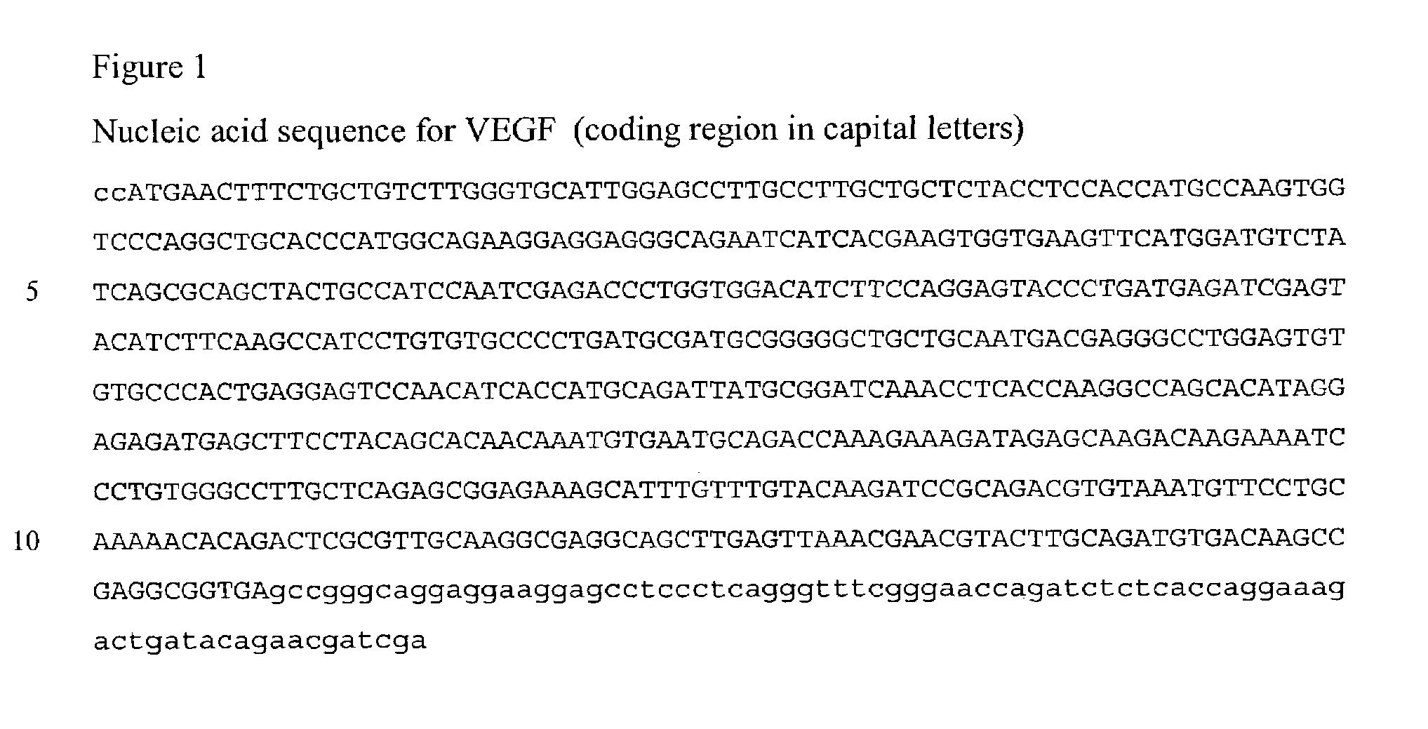
[0127]A cDNA encoding VEGF165 was isolated by PCR using primers based on published sequences (Houck, et al., Mol Endocrinol 5:1806-14. (1991)). The coding region for VEGF165 was cloned into the pTRE2 expression vector (Clontech, Palo Alto, Calif.). This vector contains a minimal CMV promoter flanked by seven repeats of the Tet operator. The integrity of the cloned VEGF-A PCR product was confirmed by restriction analysis and DNA sequencing using VEGF-specific primers.
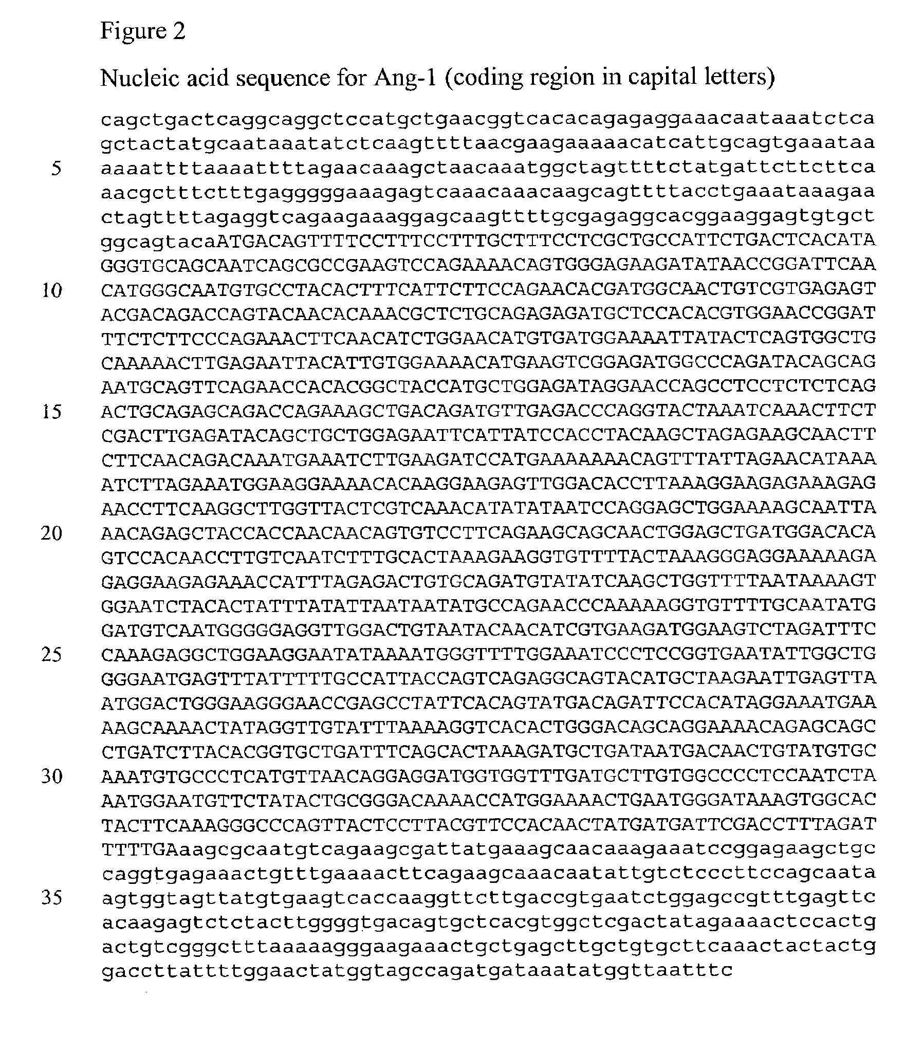
[0128]The coding region for Ang-1 was isolated by PCR from total RNA using primers derived from the published Ang-1 sequence (Maisonpierre, et al., Science 277:55-60. (1997)). The coding region for Ang-1 was cloned into the pTRE2 expression vector and the integrity of the Ang-1 coding region was verified by restriction analysis and DNA sequencing using primers derived from the known Ang-1 sequence.
[0129]The coding regions for VEGF165 and Ang-1 also were cloned into an expression vector contain...
example 3
Inducible Expression of VEGF
[0132]Purified DNA from the pTRE2 / VEGF165 vector was introduced into NIKS cells along with the pTet-On plasmid (Clontech, Palo Alto, Calif.), which encodes a derivative of the tet repressor protein. This protein, rtTA, binds to the tet operator in the presence of doxycycline and will induce expression of VEGF165 when doxycycline is present in the culture medium (see FIG. 4).
[0133]NIKS cells were transfected using TransIt-Keratinocyte reagent (Mirus Corporation). Initially, unselected populations of transfected cells were analyzed for VEGF165 expression to confirm that VEGF-A expression was induced by the presence of doxycycline. Twenty-four hours after transfection, cells were incubated with media containing doxycycline (0, 1, 10, 100, 1000 ng / ml) to induce VEGF-A expression and secretion. Media was collected 24 hr after doxycycline addition and the level of VEGF165 was determined by Western blotting using commercially-available antibodies (R&D Systems).
[...
PUM
| Property | Measurement | Unit |
|---|---|---|
| time | aaaaa | aaaaa |
| pH | aaaaa | aaaaa |
| humidity | aaaaa | aaaaa |
Abstract
Description
Claims
Application Information