Determination and uses of the atomic structures of ribosomes and ribosomal subunits and their ligand complexes

a technology of ribosomes and atomic structures, applied in the field of determination and use of ribosomes and ribosome subunits and their ligand complexes, can solve the problems of ribosomes having inherent flexibility and instability, difficult crystallization of ribosomes, and difficult acquisition of crystals, etc., and achieve the effect of sufficient quality

Inactive Publication Date: 2009-03-17
YALE UNIV
View PDF34 Cites 7 Cited by
  • Summary
  • Abstract
  • Description
  • Claims
  • Application Information

AI Technical Summary

Problems solved by technology

This was a challenging enterprise.
Crystals of ribosomes have been especially difficult to obtain because of their huge size and their lack of internal symmetry.
Moreover, since their surface is composed of highly degradable RNA and loosely held proteins, ribosomes exhibit inherent flexibility and instability.
However, the low resolution electron density maps of Schlunzen et al.
Although the prior art provides crystals of the 50S ribosomal subunit and 9 Å and 5 Å resolution X-ray crystallographic maps of the structure of the 50S ribosome, the prior art crystals and X-ray diffraction data are not sufficient to establish the 3D structures of all 31 proteins and 3,043 nucleotides of 50S.
Thus, the prior art crystals and maps are inadequate for the structure-based design of active agents, such as herbicides, drugs, insecticides, animal poisons, etc.
Each atom in a crystal scatters X-rays in all directions, but crystalline diffraction is observed only when the crystal is oriented relative to the X-ray beam so that the atomic scattering interferes constructively.
Unfortunately, the phase information essential for computing electron distributions cannot be measured directly from diffraction patterns.
In fact, however, some interference is negative; consequently, following heavy-metal substitution, some spots measurably increase in intensity, others decrease, and many show no detectable difference.
The interpretation of the electron-density map is made more complex by several limitations of the data.
First of all, the map itself contains errors, mainly due to errors in the phase angles.
Building the initial model is a trial-and-error process.
The residual difference is a consequence of errors and imperfections in the data.

Method used


Image

  • Determination and uses of the atomic structures of ribosomes and ribosomal subunits and their ligand complexes
  • Determination and uses of the atomic structures of ribosomes and ribosomal subunits and their ligand complexes
  • Determination and uses of the atomic structures of ribosomes and ribosomal subunits and their ligand complexes

Examples


specific embodiments

A. Atomic Structure of the Large Ribosomal Subunit at 2.4 Å Resolution

[0184]The present invention is based in part on the development of a novel method for preparing crystals of ribosomes. The novel method provides crystals of the 50S ribosomal subunit that are much thicker than those available earlier and that can diffract X-rays to about 2.4 Å resolution. The newly developed method eliminates the twinning of crystals that obstructed progress in determining the crystal structure of the 50S ribosomal subunit from H. marismortui for many years. The method of preparing the crystals of the 50S ribosomal subunit is discussed below.

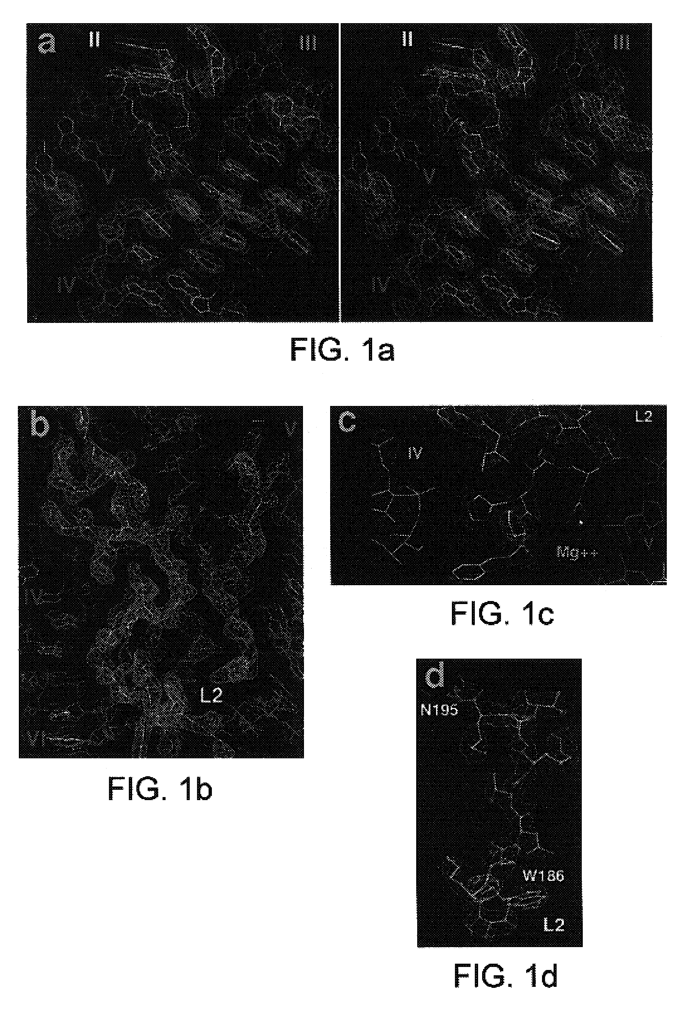
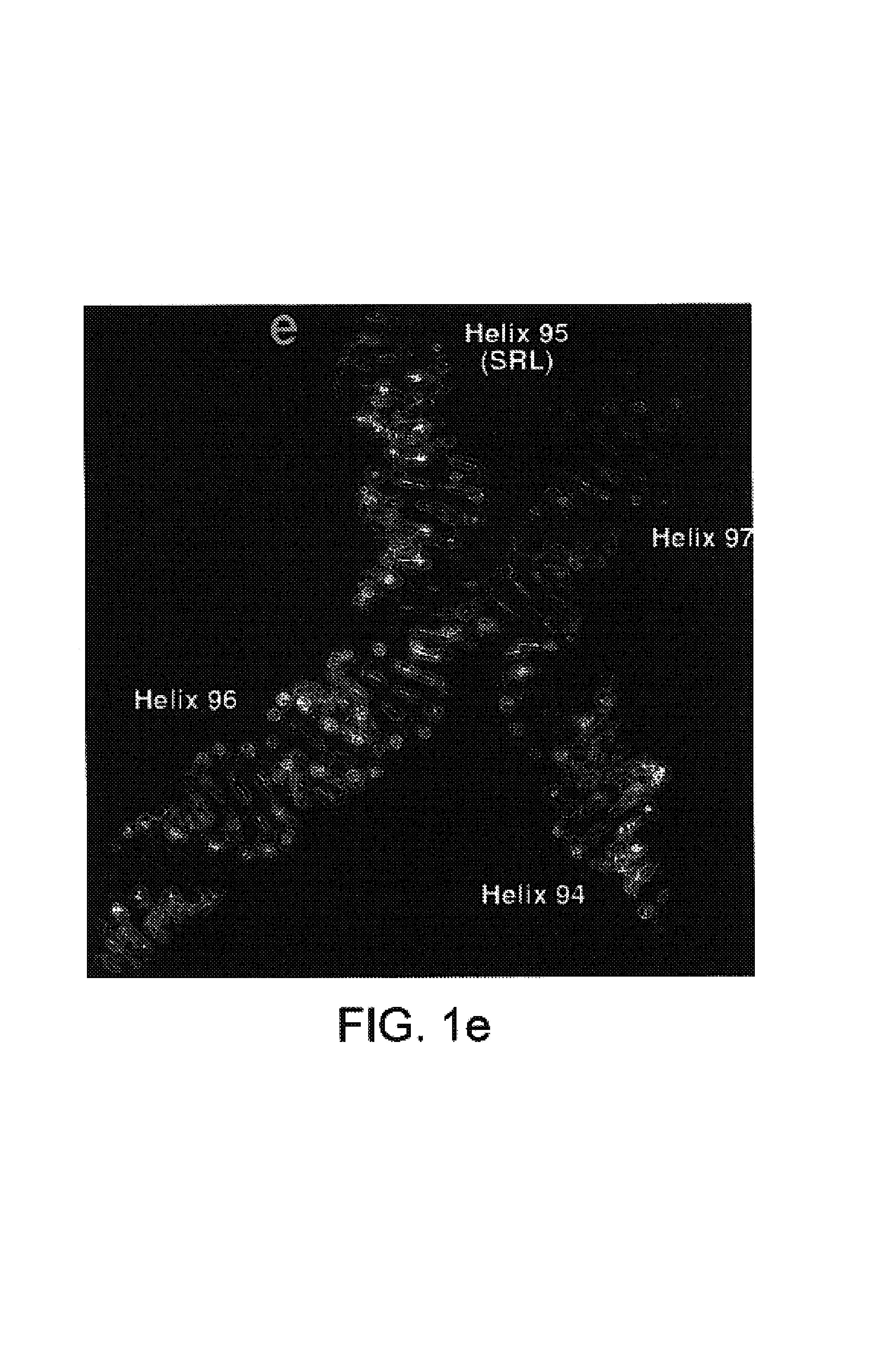
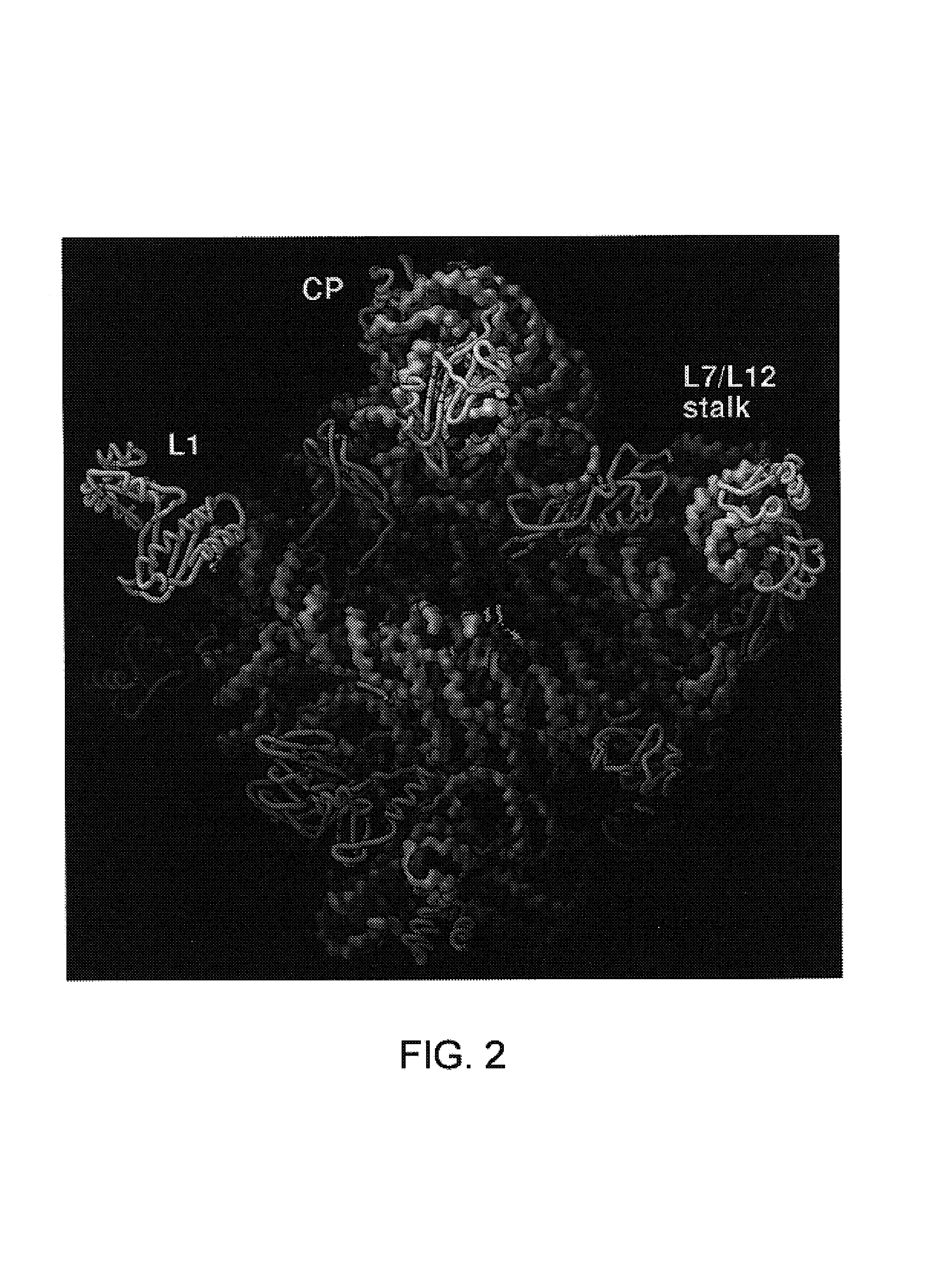
[0185]The present invention is also based in part on the atomic structure of the crystal of the 50S ribosomal subunit from H. marismortui that has been derived from a 2.4 Å resolution electron density map that was experimentally phased using heavy atom derivatives. The atomic coordinates of the structure were deposited on Jul. 10, 2000, at Research Collaborato...

example 1

Preparation of 50S Ribosomal Subunit Crystals

[0385]H. marismortui (ATCC 43049) was grown as described previously (Ban et al., 1998) on a slightly modified version of ATCC culture medium 1230, which was supplemented with 4.3 g of yeast extract, 5.1 g of Tris, and 3.4 g of glucose per liter. Bacteria were grown at 37° C. to an OD550nm between 1.0 and 2.2. They were harvested by centrifugation, and stored at −80° C. Cells were ruptured using a French press. Ribosomes were prepared from lysates by centrifugation, and subunits were isolated on sucrose gradients (Shevack et al., 1985).

Reverse Extraction

[0386]1. Take 1 mg of subunits from a concentrated 50S ribosomal subunit stock (30 mg / ml in 1.2 M KCl, 0.5 M NH4Cl, 20 mM MgCl2, Tris 10 mM, CdCl2 1 mM, Tris 5 mM, pH 7.5) and mix with ½ vol. of 30% PEG6000 (300 g PEG, 700 ml H2O to make 1 liter of 30% PEG; filter through 0.2 μm filter)

[0387]Leave on ice for 1 to 2 hr.[0388]2. Spin down precipitate ˜30 sec. using a desktop centrifuge.[0389]...

example 2

Determination of the Crystal Structure of the 50S Ribosomal Subunit

[0413]All data, except the two native data sets, were collected at the National Synchrotron Light Source (Brookhaven) from crystals frozen at 100 K, using beamlines X12b and X25 and recorded using a 345 mm MAR imaging plate. For each heavy atom derivative, anomalous diffraction data were collected at the wavelength corresponding to the peak anomalous scattering. The beam size was 100×100 μm for most data collections at X25 and 200×200 μm at beamline X12b. The crystals were aligned along the long axis of the unit cell (570 Å) so that 1.0° oscillations could be used to collect reflections out to a maximum of 2.7 Å resolution at the edge of the MAR detector. At beamline X12b the crystal to detector distances varied between 450.0 mm and 550.0 mm depending on wavelength, crystal quality and beam divergence, and it was chosen so that maximum resolution data could be collected while avoiding overlapping of spots. At beamlin...


PUM

PropertyMeasurementUnit
thicknessaaaaaaaaaa
thicknessaaaaaaaaaa
thicknessaaaaaaaaaa

Abstract

The present invention provides methods for producing high resolution crystals of ribosomes and ribosomal subunits as well as the crystals produced by such methods. The x-ray diffraction patterns of the crystals provided by the present invention are of sufficiently high resolution for determining the three-dimensional structure of ribosomes and ribosomal subunits, for identifying ligand binding sites on ribosomes and ribosomal subunits, and for molecular modeling of ligands which interact with ribosomes and ribosomal subunits. The present invention provides methods for identifying ribosome-related ligands and methods for designing ligands with specific ribosome-binding properties. Thus, the methods of the present invention may be used to produce ligands which are designed to kill or inhibit any specific target organism(s).

Description

RELATED APPLICATIONS[0001]This application is a divisional of U.S. Serial No. 09 / 635,708, filed Aug. 9, 2000, which issued as U.S. Pat. No. 6,638,908 B1 on Oct. 28, 2003, the entire disclosure of which is incorporated by reference herein.ACKNOWLEDGMENT OF FEDERAL SUPPORT[0002]This work was at least partially funded by the following NIH grants: NIH-GM22778 and NIH-GM54216.FIELD OF THE INVENTION[0003]The present invention generally pertains to the fields of molecular biology, protein crystallization, x-ray diffraction analysis, three-dimensional structural determination, structure based rational drug design and molecular modeling of ribosomes and ribosomal subunits. The present invention provides crystallization methods as well as the crystallized ribosome and ribosomal subunits. The x-ray diffraction patterns of the crystals in question are of sufficient resolution so that the three-dimensional structure of ribosomes and ribosomal subunits can be determined at atomic resolution, liga...

Claims


Application Information

Patent Timeline
Patent Type & Authority Patents(United States)
IPC IPC(8): C07K1/14G01N31/00C07K14/215
CPCC07K14/215C07K2299/00
Inventor STEITZ, THOMAS A.MOORE, PETER B.BAN, NENADNISSEN, POULHANSEN, JEFFREY
Owner YALE UNIV
Why Eureka
Try Eureka