

Isolated peptides to treat alpha-synuclein diseases
a technology of alpha-synuclein and isolated peptides, which is applied in the direction of peptide/protein ingredients, peptide sources, instruments, etc., can solve the problems of affecting the development of screening assays, affecting the aggregation of -synuclein in living neurons, and oxidative conditions that actually promote -synuclein aggregation in living neurons
- Summary
- Abstract
- Description
- Claims
- Application Information
AI Technical Summary
Problems solved by technology
Method used
Image

Examples
example 1
Iron and Free Radicals Stimulate α-Synuclein Aggregation
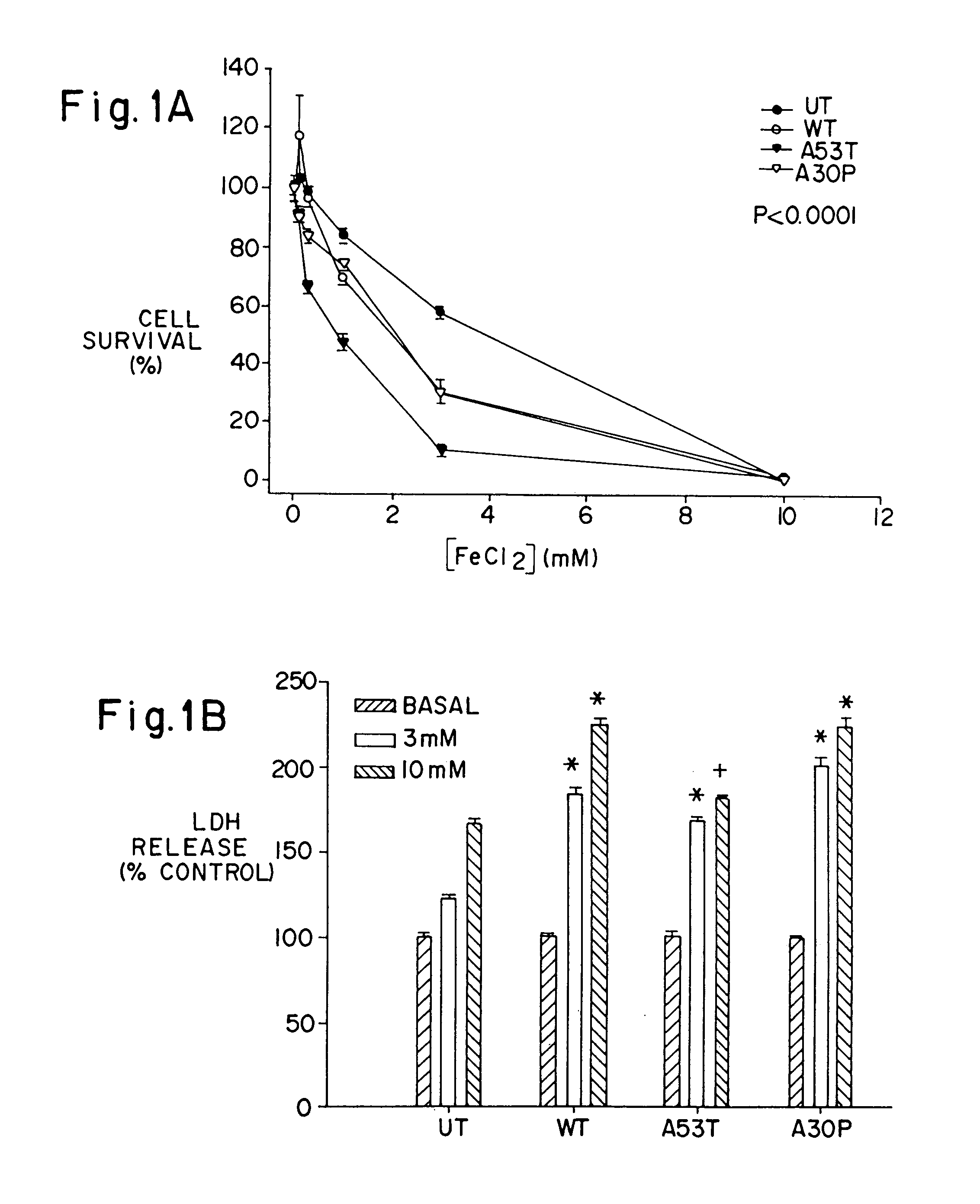
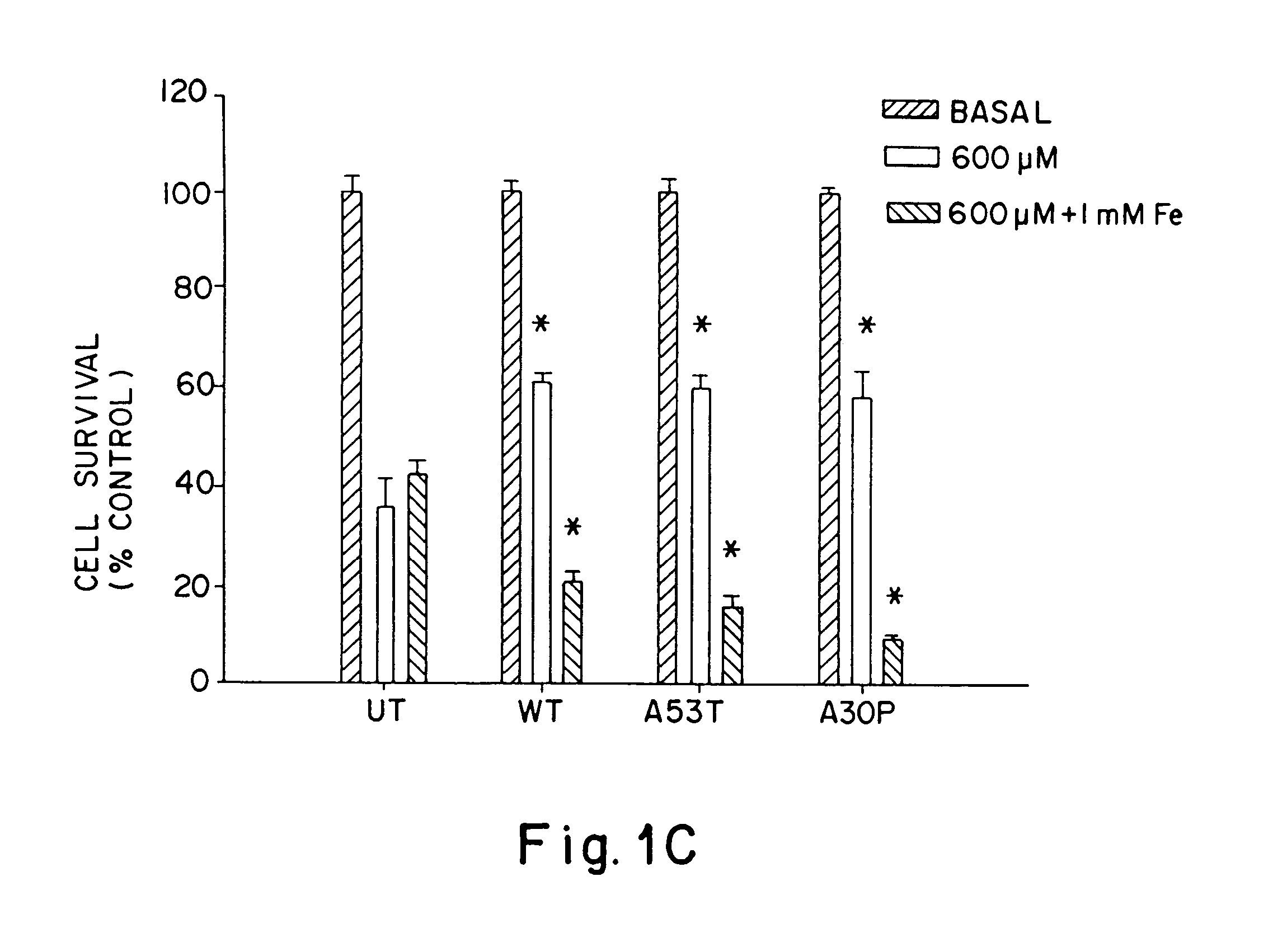
[0064]To test whether A53T and A30P mutations in α-synuclein increase the tendency of α-synuclein to aggregate in neurons, α-synuclein aggregation was determined in human BE-M17 neuroblastoma cells that were stably transfected with wild type, A53T or A30P α-synuclein. Each cell line was treated for 48 hours with freshly prepared FeCl2 (1 or 10 mM), then harvested, homogenized and fractionated into membrane and cytoplasmic components. The membrane (5 μg / lane) and cytoplasmic (20 μg / lane) components were immunoblotted with monoclonal anti-α-synuclein antibody.
[0065]Aggregates of α-synuclein were evident in the membrane fraction, but not in the cytoplasmic fraction. Treatment of the A53T-expressing cell line with FeCl2 induced dose-dependent formation of heterogeneous high molecular weight-α-synuclein aggregates that migrated in the stacking gel. A large amount of anti-α-synuclein immunoreactivity was also apparent in the upper po...
example 2
A53T and A30P α-Synuclein Aggregates Contain Ubiguitin
[0070]In PD, Lewy bodies have also been shown to contain large amounts of ubiquitin [Dickson, D., et al., Brain Path., 9:721-32 (1999); Gibb, W., et al., J. Neurol. Neurosurg. and Psych. 51:745-52 (1988)]. To determine whether aggregation of ubiquitin also occurred along with α-synuclein aggregation, lysates (membrane fractions) were taken from the BE-M17 cells expressing A30P α-synuclein that had been treated with FeCl2 and dopamine (see Example 1, 48 hr treatment), and were immunoblotted with anti-ubiquitin antibody. The ubiquitin aggregates that accumulated under these conditions stained strongly for ubiquitin. Aggregates that accumulated in cells expressing A53T α-synuclein or wild type α-synuclein after being treated with either FeCl2 alone, FeCl2 plus hydrogen peroxide or FeCl2 plus dopamine also contained ubiquitin. The amount of aggregated ubiquitin generally paralleled the amount of α-synuclein. The presence of ubiquitin...
example 3
α-Synuclein Aggregates Form Visible Inclusions Evident by Thioflavine-S Histochemistry and Electron Microscopy
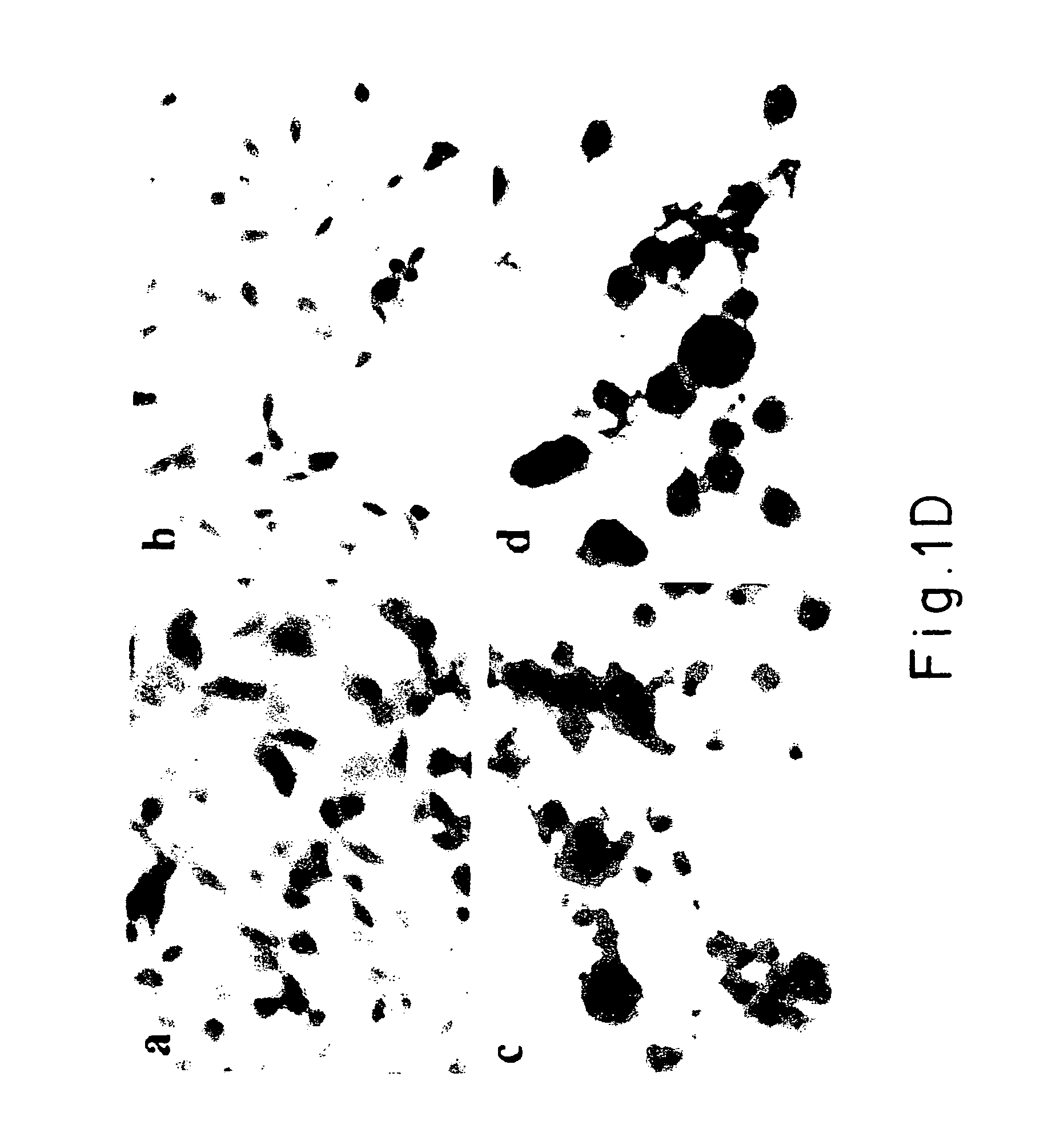
[0071]Thioflavine-S histochemistry and electron microscopy were used to examine the aggregates that formed in response to treatment with iron and hydrogen peroxide. Cells from each line (BE-M17: untransfected, wild type, A30P and A53T a.-synuclein) were treated with 10 mM FeCl2 or 10 mM FeCl2 plus 100 μM H2O2 for 72 hrs to induce formation of α-synuclein positive inclusions. The aggregates that formed were observed to bind thioflavine-S, which indicates the presence of β-pleated sheet structures. The size and number of thioflavine-S-positive aggregates paralleled the results seen by immunoblotting. The wildtype, A30P and A53T α-synuclein cell lines each showed significant accumulations of protein aggregates after treatment for 72 hrs with. 10 mM FeCl2 plus 100 μM H2O2. In contrast, the untransfected line showed no accumulation of thioflavine-S positive aggregates. When only ...
PUM
| Property | Measurement | Unit |
|---|---|---|
| diameter | aaaaa | aaaaa |
| time | aaaaa | aaaaa |
| pH | aaaaa | aaaaa |
Abstract
Description
Claims
Application Information