
Endoscope retrieval instrument assembly

a technology for removing instruments and endoscopes, applied in the field of surgical instruments, can solve the problems of preventing visual inspection of the internal tissues of patients, and often becoming difficult to capture the polyp and retrieve it from the patien

Active Publication Date: 2009-11-17
GRANIT MEDICAL INNOVATION
View PDF10 Cites 90 Cited by
  • Summary
  • Abstract
  • Description
  • Claims
  • Application Information

AI Technical Summary

Benefits of technology

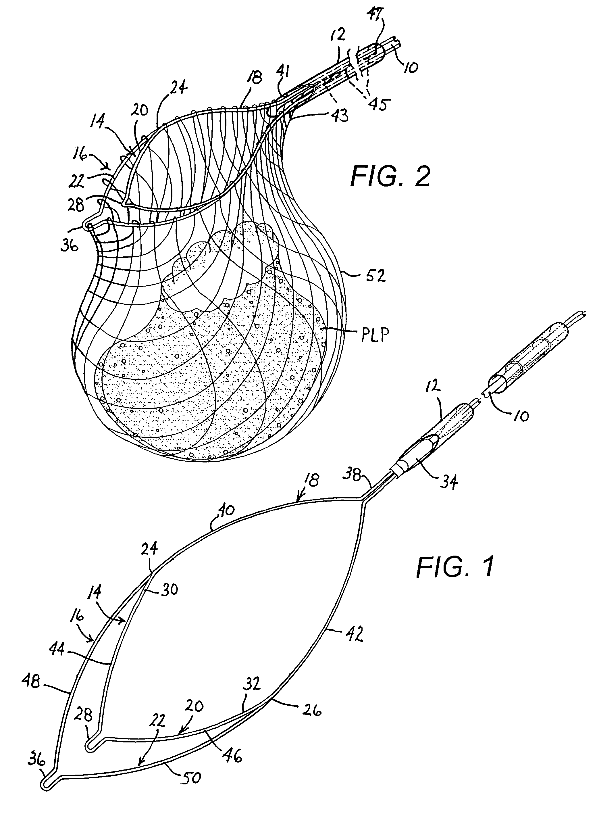
"The present invention is about a medical instrument that can be used to capture and remove tissue mass, such as polyps, from an internal organ. The instrument includes an elongate tubular introducer member, an elongate slider member, a loop, and a pouch. The instrument can be used in endoscopic interventions and includes a tether that allows the pouch to slide along at least a portion of the loop. The instrument can also include a cauterization snare with a pouch attached for tissue retrieval purposes. The invention allows for proper specimen retrieval and visualization during the intervention."

Problems solved by technology

Once a polyp is severed by such a snare cauterization technique, it frequently becomes difficult to capture the polyp and retrieve it from the patient.
The use of suction has the drawback of frequently preventing visual inspection of the patient's internal tissues during withdrawal of the endoscope.
Especially in cases where the polyp is large, the effort and time expended in retrieving the severed polyp may rival or even exceed the effort and time required to locate and sever the polyp.
Furthermore, there are numerous cases where a severed polyp is never recovered.
In all such cases, the pathologist is unable to determine whether the polyp contains carcinoma in situ (localized to the mucosa) or infiltrative carcinoma (spread beyond the muscularis mucosa layer).
In any event, the manipulations necessary to remove a severed polyp generally increase the trauma to the patient, the expense of the surgery and the hospitalization time.

Method used


Image

  • Endoscope retrieval instrument assembly
  • Endoscope retrieval instrument assembly
  • Endoscope retrieval instrument assembly

Examples


Embodiment Construction

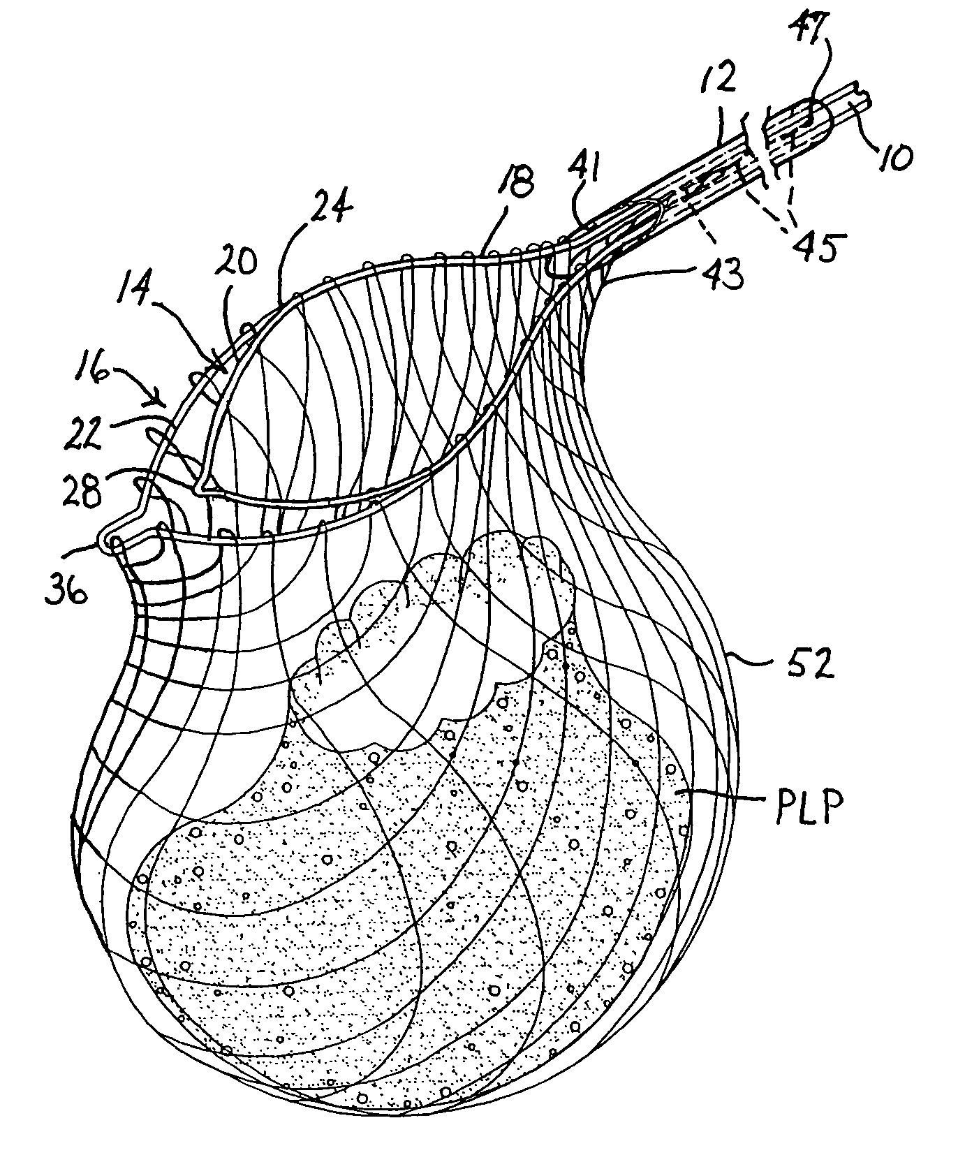
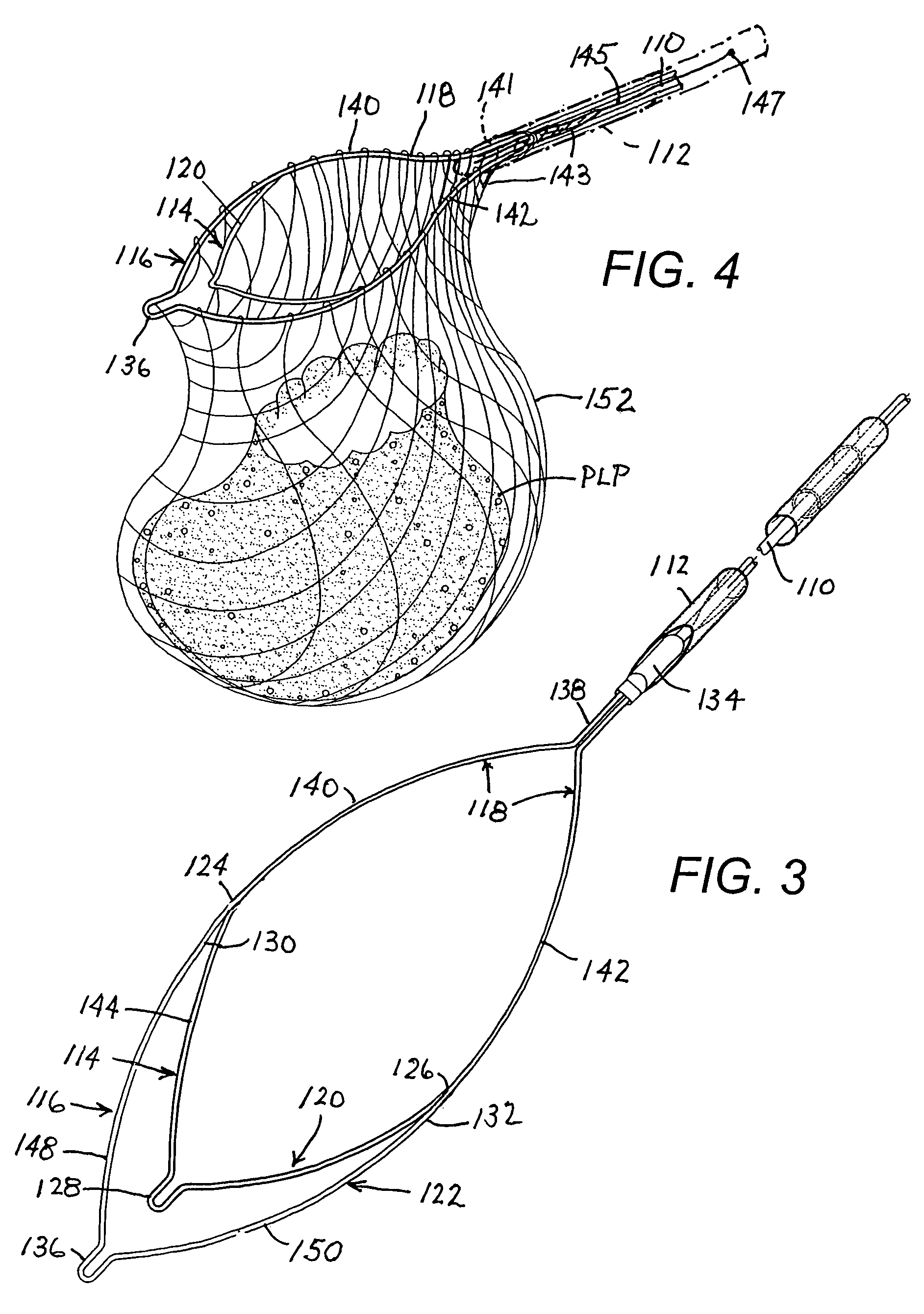
[0043]The drawings illustrate a number of different medical retrieval devices, all having a net element forming a pouch where the net element may be at least partially slidably secured along a wire loop, particularly along a proximal end portion of the loop, facing the user of the instrument. The wire loop and the pouch are secured to a distal end of a wire pusher that extends through a tubular introducer member (e.g., catheter) transmits axial motion to the loop and net element. At the onset of an endoscopic procedure the wire loop and the pouch or net element are completely housed in a tubular introducer member such as a catheter. The catheter is inserted into a patient via a working channel of an endoscope. When the distal end of the endoscope has reached a surgical or diagnostic site inside the patient, such as a polyp in the colon, the wire loop and the net element or pouch are ejected from the catheter. The wire loop automatically opens to form a mouth of the pouch and is mani...


PUM


Abstract

A medical instrument includes an elongate tubular introducer member, an elongate slider member disposed at least partially inside the tubular member, a loop provided at a distal end of the slider member, a pouch connected to the loop so that the loop defines a mouth opening of the pouch and so that the pouch is slidable along at least a proximal portion of the loop, and a tether connected to the pouch at a proximal side thereof. The tether extends into the tubular member and is fastened to the tubular member at a location spaced by a distance from a distal tip of the tubular member. The tether has a length from the fastening location to the pouch that is less than the distance between the location and the tip of the tubular member.

Description

FIELD OF THE INVENTION[0001]This invention relates to a surgical instrument assembly for use in retrieving objects from internal body cavities. This invention also relates, more specifically, but not exclusively, to a surgical instrument assembly for use in snare cauterization operations.BACKGROUND OF THE INVENTION[0002]In a conventional endoscopic snare operation, an endoscope is inserted into an internal cavity of a patient, e.g., into the colon, and is used to locate abnormal tissue growths such as polyps in the internal cavity. Upon the locating of a polyp or other growth which is to be removed, a wire extending through a tube in the biopsy channel of the endoscope is slid in the distal direction so that a cauterization loop connected to the wire is ejected from the distal end of the tube and the endoscope. The loop and the endoscope are manipulated from outside of the patient to pass the loop over the polyp or growth. The wire is then withdrawn in the proximal direction to tigh...

Claims


Application Information

Patent Timeline
Patent Type & Authority Patents(United States)
IPC IPC(8): A61B17/26
CPCA61B17/221A61B17/32056A61B2017/2212A61B2017/00287A61B2017/00269
Inventor NAKAO, NAOMI L.
Owner GRANIT MEDICAL INNOVATION
Learn More