
Linking of images to enable simultaneous viewing of multiple objects

a technology of linking images and objects, applied in image enhancement, fluid pressure computing, instruments, etc., can solve the problems of poor and/or long-repetitive analysis, relatively delicate serial sections, and add to cross-comparing serial sections difficulty, so as to increase or decrease the transparency of ghost images

Active Publication Date: 2009-11-24
LEICA BIOSYST IMAGING
View PDF3 Cites 28 Cited by
  • Summary
  • Abstract
  • Description
  • Claims
  • Application Information

AI Technical Summary

Benefits of technology

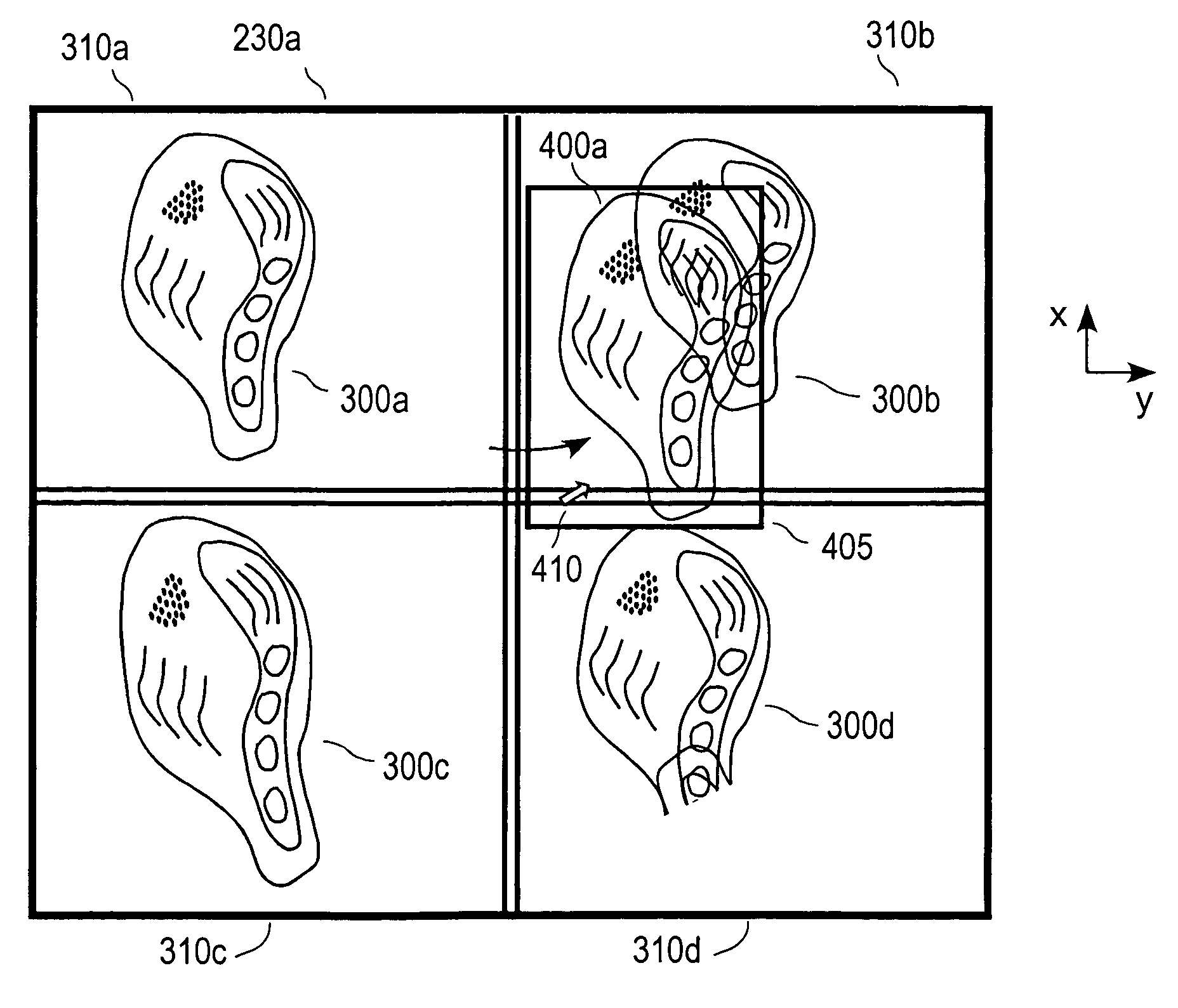
[0008]In short, this is made possible by the use of a computerized system and computerized techniques for linking the digitized images of the serial sections. In a method of linking digitized images of serial sections, according to one embodiment, the digitized images of the serial sections are displayed on a computer display that is controlled by a computer, a copy of a digitized image of a first serial section is created, and the copy is referred to as the ghost image or transparent image. The ghost image is positioned over a digitized image of ...

Problems solved by technology

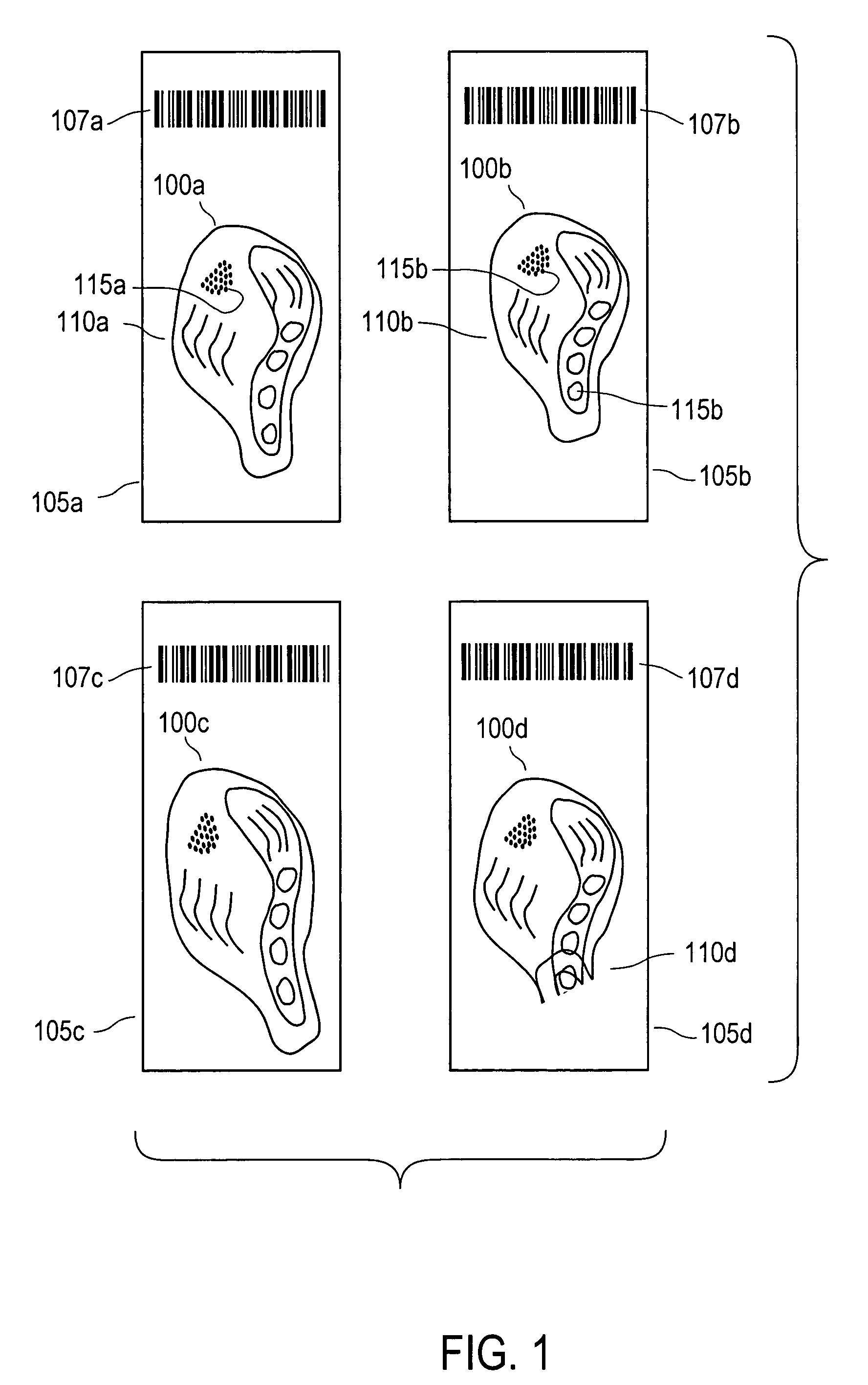
Being relatively thin, the serial sections are relatively delicate and when placed on microscope slides tend to deform by stretching, shrinking, being compressed, folding or a combination thereof.
Such deformations and relative rotations often add to the difficulty in cross comparing serial sections.
As corresponding tissues may be distorted compared to the H&E section, and / or in a different location or orientation, position estimates may be many millimeters off leading to poor and / or lengthy-repetitious analysis.
Poor and lengthy analysis arise not only in naked eye analysis of serial sections but also in computer-aided analysis of serial sections.
Present computer-aided analysis techniques do not correct for distortions and relative rotations of serial sections captured in digital images of these sections.
As a result of the distortion and relative rotations of a set of serial images captured in digitized images, using location information derived from one serial-section image to locate structures in another serial-section image using computer-aided techniques is a laborious process fraught with misidentification and lengthy, repetitious analysis.

Method used


Image

  • Linking of images to enable simultaneous viewing of multiple objects
  • Linking of images to enable simultaneous viewing of multiple objects
  • Linking of images to enable simultaneous viewing of multiple objects

Examples


Embodiment Construction

Overview

[0034]The present invention provides a system and a technique for using the system for cross-comparing and analyzing cross sections (or “serial sections”) of biological tissue samples, such as tumorous tissues, using a computer to correct for deformations of the serial sections captured in digitized images. Systems and techniques are also provided for correcting relative rotational displacement of the digitized images and for linking the digitized images, such that graphical manipulations performed on one serial image are similarly performed on linked serial images.

[0035]A particular application of the present invention is in the field of pathology, and other medical or bioscience fields, to correct for distortion and relative rotations between digitized images of serial sections variously stained to color select structures of interest in the serial sections. A first serial section of a tissue sample, often used as a reference section, is typically stained with haematoxylin ...


PUM


Abstract

Computerized techniques are provided for linking digitized images of serial sections of a biological tissue sample. Linked, digitized images of the serial section are displayed on a display and manipulation of one digitized image causes other digitized images to be similarly manipulated. To link digitized images, a copy of a digitized image of one serial section is positioned over a digitized image of another serial section. The digitized image of the other serial section is visible through the copy. The transparency of the copy is changed to effect change of the visualization of the digitized image of the other serial section. The copy is registered to the digitized image of the other serial section.

Description

BACKGROUND OF THE INVENTION[0001]This invention relates generally to the analysis of samples such as biological tissue samples that are chemically stained for protein based markers, and more specifically to the use of a linking system and method for linking sets of images of the samples in such analysis.[0002]Visual analysis of biological tissue samples often involves slicing the biological tissue samples into thin cross sections, often referred to as serial sections, to visualize structures of interest within the biological tissue sample. The serial sections are typically mounted on glass or plastic microscope slides. Visual analysis of mounted serial sections is often carried out by the naked eye (grossly) or by microscopy. In a typical slicing process, a tissue sample is dehydrated and embedded in paraffin to lend rigidity to the sample during slicing and mounting on slides. Tissue samples are typically sliced into serial sections that are about 4-9 micrometers (μm) thick; howeve...

Claims


Application Information

Patent Timeline
IPC IPC(8): G06K9/00G06G5/00
CPCG06F3/04845G06T7/0024G06T2207/30024G06T2207/10056G06T2207/20092G06T2200/24G06T7/30
Inventor HUGHES, WILLIAMBORTHWICK, DUNCAN WILLIAM
Owner LEICA BIOSYST IMAGING
Learn More