System and method for ECG-triggered retrospective color flow ultrasound imaging

a color flow ultrasound and imaging system technology, applied in tomography, instruments, applications, etc., can solve the problems of limited ability to detect low flow rate, spatio-temporal decorrelation artifacts that occur, and limited flow velocity estimation accuracy

Active Publication Date: 2010-03-09
SUNNYBROOK & WOMENS COLLEGE HEALTH SCI CENT +1
View PDF129 Cites 44 Cited by
  • Summary
  • Abstract
  • Description
  • Claims
  • Application Information

AI Technical Summary

Problems solved by technology

This technique, however, has limitations including tissue clutter artifacts that are induced by the sweep velocity, which limits the ability to detect low flow rates.
Other limitations include spatio-temporal decorrelation artifacts that occur when visualizing pulsatile flow, particularly if the pulse frequency is large relative to the sweep frequency of the probe.
Moreover, an additional limitation includes limited accuracy of flow velocity estimation because of the number of radio frequency (RF) data lines acquired per location.

Method used


Image

  • System and method for ECG-triggered retrospective color flow ultrasound imaging
  • System and method for ECG-triggered retrospective color flow ultrasound imaging
  • System and method for ECG-triggered retrospective color flow ultrasound imaging

Examples


example 1

In Vivo Carotid Imaging Using ECG-triggered Retrospective Color Flow Imaging

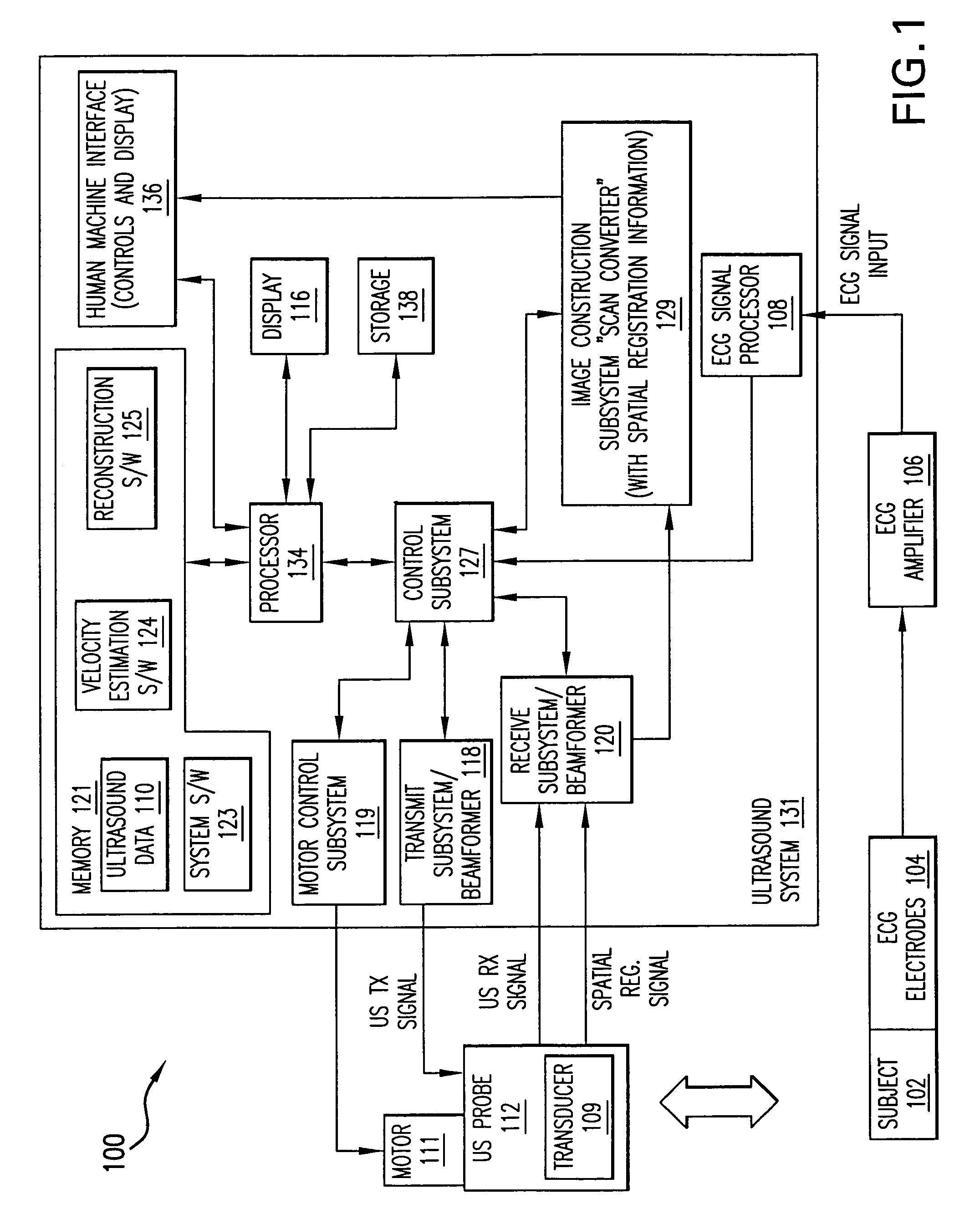
[0065]For swept-scan data acquisition, a Vevo660 ultrasound biomicroscope (UBM) system 802 (FIG. 8)(Visualsonics, Toronto, ON, Canada) was used to transmit and receive ultrasound data. The system was set to generate seven cycle pulses by internally gating and amplifying the CW signal produced by a master oscillator 804.

[0066]For in vivo carotid imaging, 40 MHz pulses were transmitted by an ultrasound probe 112 with a transducer 109. For example, an RMV604 probe equipped with a 40 MHz transducer (6 mm focal length) at a PRF of 10 kHz was used. For color flow imaging, received signals were demodulated using a demodulating element 806 by the Vevo660 802 using the CW signal from its master oscillator 804 to produce in-phase (I) and quadrature-phase (Q) signals that were digitized by an analog to digital converter (A / D) 808.

[0067]Transmitted pulses were generated using the CW signal provided by the master oscilla...

example 2

In Vitro EGC Retrospective Color Flow Imaging Using a Phantom

[0075]Both swept-scan color flow imaging and ECG-triggered retrospective color flow imaging were compared using a phantom with a 5-Hz sinusoidally varying velocity profile. The phantom comprises an off-center rotating disk, with an optical sensor which generates an ECG-like pulses on each rotation of the disk.

[0076]With a swept-scan technique, good estimation of velocities between 4 mm / s and 35 mm / s were achieved, while with the retrospective technique as described above, good estimation of velocities between 2 mm / s and 35 mm / s were achieved. Spatio-temporal decorrelation artifacts were also examined for each technique. Multiple frames of the swept-scan color flow mapping showed that the locations of velocity components were incoherently positioned between frames, with a frame-rate dependent on the sweep frequency. Multiple frames of the ECG-triggered retrospective color flow mapping, however, showed a gradual velocity cha...


PUM


Abstract

A method for producing an ECG-triggered retrospective color-flow ultrasound image comprises generating ultrasound, transmitting the ultrasound into a subject at a first location, wherein a first reference point of an ECG signal taken from the subject triggers the ultrasound transmission, receiving ultrasound reflected from the subject at the first location, transmitting the ultrasound into the subject at a second location, wherein a second reference point of an ECG signal taken from the subject triggers the ultrasound transmission receiving ultrasound reflected from the subject at the second location, processing the received ultrasound to form ultrasound color traces, and reconstructing the ultrasound color traces to form the ultrasound image.

Description

CROSS-REFERENCE TO RELATED APPLICATIONS[0001]This application claims the benefit of U.S. Provisional Application No. 60 / 549,041, filed on Mar. 1, 2004. The aforementioned application is herein incorporated by reference in its entirety.Names of the Parties to a Joint Research Agreement[0002]Sunnybrook and Women's College Health Sciences Centre and VisualSonics Inc. are parties to a joint research agreement.BACKGROUND OF INVENTION[0003]Small animal or laboratory animal research is a cornerstone of modem biomedical advancement. Research using small animals enables researchers to understand complex biological mechanisms, to understand human and animal disease progression, and to develop new drugs to cure or alleviate many human and animal maladies. Small animal research is important in many areas of biomedical research including neurobiology, developmental biology, cardiovascular research and cancer biology.[0004]In many areas of biomedical research, accurately determining blood flow ch...

Claims


Application Information

Patent Timeline
Patent Type & Authority Patents(United States)
IPC IPC(8): A61B8/00G06K9/00A61B8/06G01S7/52G01S15/89
CPCA61B8/06A61B8/13A61B8/5284A61B8/54G01S7/52034G01S7/52085G01S7/52087G01S7/52088G01S15/8906G01S15/8956G01S15/8988G01S15/899G01S15/8979
Inventor WILLIAMS, ROSSNEEDLES, ANDREWCHERIN, EMMANUELFOSTER, F. STUART
Owner SUNNYBROOK & WOMENS COLLEGE HEALTH SCI CENT
Why Eureka
Try Eureka