Surgical imaging device

a surgical imaging and device technology, applied in the field of surgical imaging devices, can solve the problems of surgeons not being able to see the surgical site, difficult to maneuver, single view of the surgical site,

Inactive Publication Date: 2010-07-06
TYCO HEALTHCARE GRP LP
View PDF180 Cites 1171 Cited by
  • Summary
  • Abstract
  • Description
  • Claims
  • Application Information

AI Technical Summary

Benefits of technology

[0006]In accordance with one example embodiment of the present invention, a surgical imaging device is provided. The surgical imaging device is configured to be inserted into a surgical site. The surgical imaging device includes a plurality of prongs. Each one of the prongs has an image sensor mounted thereon. The image sensors provide different image data corresponding to the surgical site, thus enabling a surgeon to view a surgical site from several different angles.

Problems solved by technology

However, there are many types of surgical procedures in which the surgeon is not able to see the surgical site.
However, once one of these cameras is inserted through an incision and into a surgical site, it may be difficult to maneuver due to the fact that it is prevented from movement in most directions by the small incision.
In addition, these cameras provide only a single view of the surgical site.

Method used


Image

  • Surgical imaging device
  • Surgical imaging device
  • Surgical imaging device

Examples


Embodiment Construction

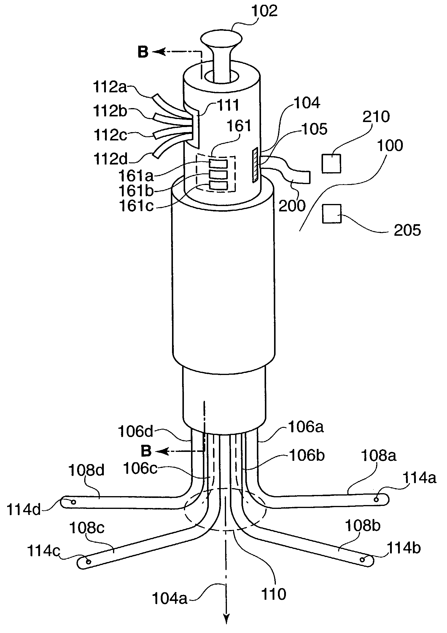
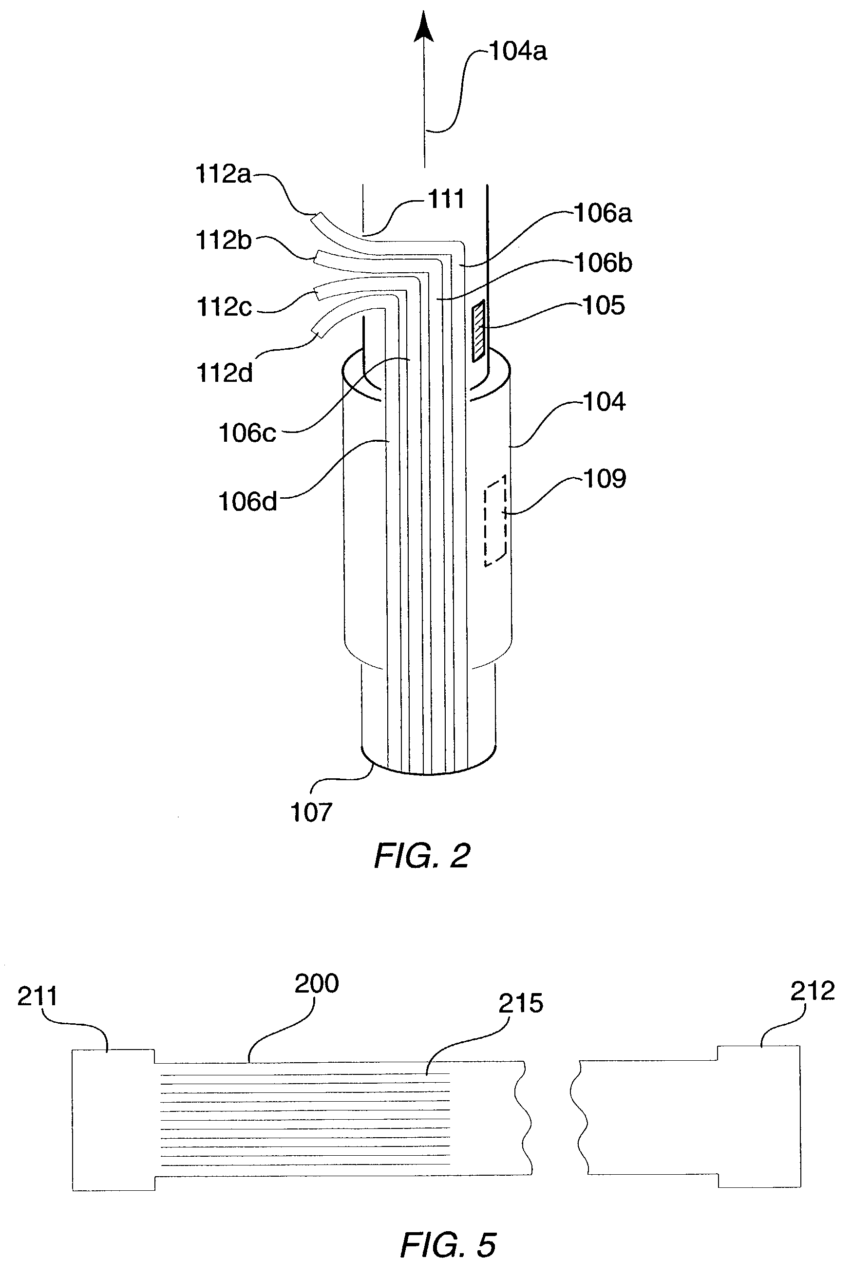
[0022]FIG. 1 shows a perspective view of a surgical imaging device 100 according to an example embodiment of the present invention. The surgical imaging device 100 includes a body portion 104 which encloses legs 106a to 106d and a retraction actuator 102. The legs 106a to 106d are connected to levers 112a to 112d, respectively. Prongs 108a to 108d extend from legs 106a to 106d, respectively. Located at or near the distal tip of each prong 108a to 108d is a camera 114a to 114d, respectively.

[0023]According to one embodiment of the present invention, the legs 106a to 106d, along with their respective prongs 108a to 108d, are moveable. For instance, the legs 106a to 106d may be moveable within a cylindrical opening of the body portion 104 (explained in more detail below) so that the legs 106a to 106d move radially around a central axis 104a of the body portion 104. In addition, the legs 106a to 106d may be rotatably moveable, e.g., rotatable around their own central axes, within the bo...


PUM


Abstract

A surgical imaging device and method configured to be inserted into a surgical site. The surgical imaging device includes a plurality of prongs. Each one of the prongs has an image sensor mounted thereon. The image sensors provide different image data corresponding to the surgical site, thus enabling a surgeon to view a surgical site from several different angles. The prongs may be moveable between a first position, suitable for insertion though a small surgical incision, and a second position, in which the prongs are separated from each other. In addition, the prongs may be bendable.

Description

CROSS-REFERENCE TO RELATED APPLICATIONS[0001]This application claims the benefit under 35 U.S.C. §119(e) of U.S. Provisional Patent Application No. 60 / 352,726, filed on Jan. 30, 2002, which is expressly incorporated herein by reference in its entirety.FIELD OF THE INVENTION[0002]The present invention relates to a surgical imaging device, and in particular to a surgical imaging device that is configured to provide image data of a body cavity.BACKGROUND INFORMATION[0003]It is typically important during a surgical procedure that a surgeon be able to view the surgical site so as to ensure that the surgical procedure is being performed correctly. However, there are many types of surgical procedures in which the surgeon is not able to see the surgical site. For instance, laparoscopic or endoscopic surgical procedures, in which a surgeon accesses a surgical site through very small incisions, prevent the surgeon from viewing the surgical site.[0004]One method for performing surgical procedu...

Claims


Application Information

Patent Timeline
Patent Type & Authority Patents(United States)
IPC IPC(8): A61B6/00A61B19/00A61B1/00A61B1/313A61B17/00
CPCA61B1/313A61B1/00045A61B19/5212A61B1/3132A61B1/07A61B1/0669A61B1/042A61B1/00147A61B1/00114A61B19/5225A61B1/00025A61B1/00018A61B1/00016A61B1/00009A61B19/5202A61B17/00234A61B2017/003A61B2019/5217A61B2090/3614A61B90/30A61B90/361A61B90/37A61B1/00148
Inventor WHITMAN, MICHAEL P.
Owner TYCO HEALTHCARE GRP LP
Why Eureka
Try Eureka