
Medical device

a medical device and electrode technology, applied in the field of medical devices, can solve the problems of inability to maintain the moisture level needed for optimum wound healing, failure of normal healing process, and lack of appropriate electrical signals at the site,

Inactive Publication Date: 2008-05-29
ARGENTUM MEDICAL
View PDF35 Cites 64 Cited by
  • Summary
  • Abstract
  • Description
  • Claims
  • Application Information

AI Technical Summary

Benefits of technology

[0037]In certain aspects, the disclosed compositions can aid in restoring the transepithelial skin potential, maintain a moist wound healing environment, create a functional microbial barrier, reduce microbial bio-burden of the wound, deliver an antimicrobial metal, aid in reducing pain, or a combination thereof. For example, certain embodiments of the disclosed devices can reduce the wound potential or make the wound potential more negative by about 1 mV, about 5 mV, about 10 mV, about 20 mV, about 30 mV, about 40 mV, about 50 mV or more.
[0044]An additional aspect of the disclosure relates to wound dressings that provide for a capacitive effect formed by the alternation of conductive layers of fiber with non-conductive layers.

Problems solved by technology

Conversely, failure of the normal healing process, for example as in fracture non-unions, is associated with, among other things, the absence of appropriate electrical signals at the site of the injury or infection.
If the wound dressing dries out, the needed moisture level for optimum wound healing will not be maintained and the dressing will stick to the wound surface and cause disruption of cellular processes.
The lack of moisture often results in the formation of an eschar or scab, and a general slowing of the wound healing process.
Wounds that produce an extensive amount of moisture are thought to create another problem called skin maceration.
It is known to cause a breakdown of the cornified epithelium, thereby reducing the physical microbial barrier function as well as the moisture regulation function of the epidermis.
With a reduction of the microbial barrier function, the wound surface has a significantly greater risk of contamination by pathogenic microbes from the surrounding environment.
For example, in certain perforated films, the perforations are sufficient to permit wound exudates to diffuse through the film at a rate that precludes pooling on the wound surface, which is a common cause of maceration.
While there are numerous dressings designed to retain the moisture content of wounds, there are still many areas of inefficiency in current treatment methods.
For example, these dressings are only effective for moist wounds and do not provide any significant benefit for dry wounds.
In either situation, removal of the dressing can cause disruption of the cellular process of the wound and increase the risk of contamination by microbes.
Wounds cause destruction of the cornified epithelium as well as deeper layers thereto, and the loss of the natural anti-microbial barrier.
Most silver compounds that dissociate readily and produce large numbers of free silver ions are highly toxic to mammalian tissues.
Less-toxic compounds, including silver sulfadiazine cream, widely used in the treatment of burns, do not dissociate readily and therefore do not release large numbers of silver ions.
However, the technique provides a flat plating pattern and does not uniformly coat the entire three-dimensional surface of fibers.
This does not allow for direct contact of the maximum number of ions with the wound.
This requires an external power source and stationary equipment and is cumbersome for the patient.
Foils do not provide for circulation of air, and are limited in surface area.
Such compositions have a high volume resistance and very poor conductivity.
Regardless of whether silver is provided in the form of silver ions or as a topical composition (silver nitrate solution, silver sulfadiazine cream, or the like), its beneficial effects are manifested primarily at the treated surface and immediately adjacent tissues, and are limited by the achievable tissue concentration of silver ions.
None of the available metallic ion treatment devices provide an efficient and convenient means to restore the homeostatic electromagnetic environment for areas of wounds.
They also do not provide for maximum surface area for release of metallic ions.
In addition, the prior art does not address the need to regulate the moisture content of a wound without manually changing the dressings, or applying liquids or medicants.
Such activities allow for microbe contamination and interrupt the healing process.
Problems associated with applying a negative pressure to a wound include: tissue growth into the dressing; potential damage of delicate structures such as blood vessels and internal organs and adhesion of the dressing to the wound base causing repeated trauma (therefore increasing pain and increased healing time) with dressing changes.

Method used

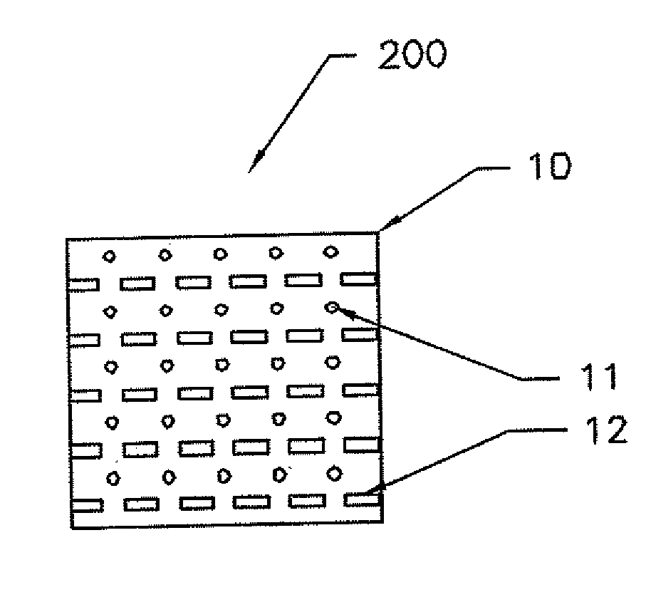
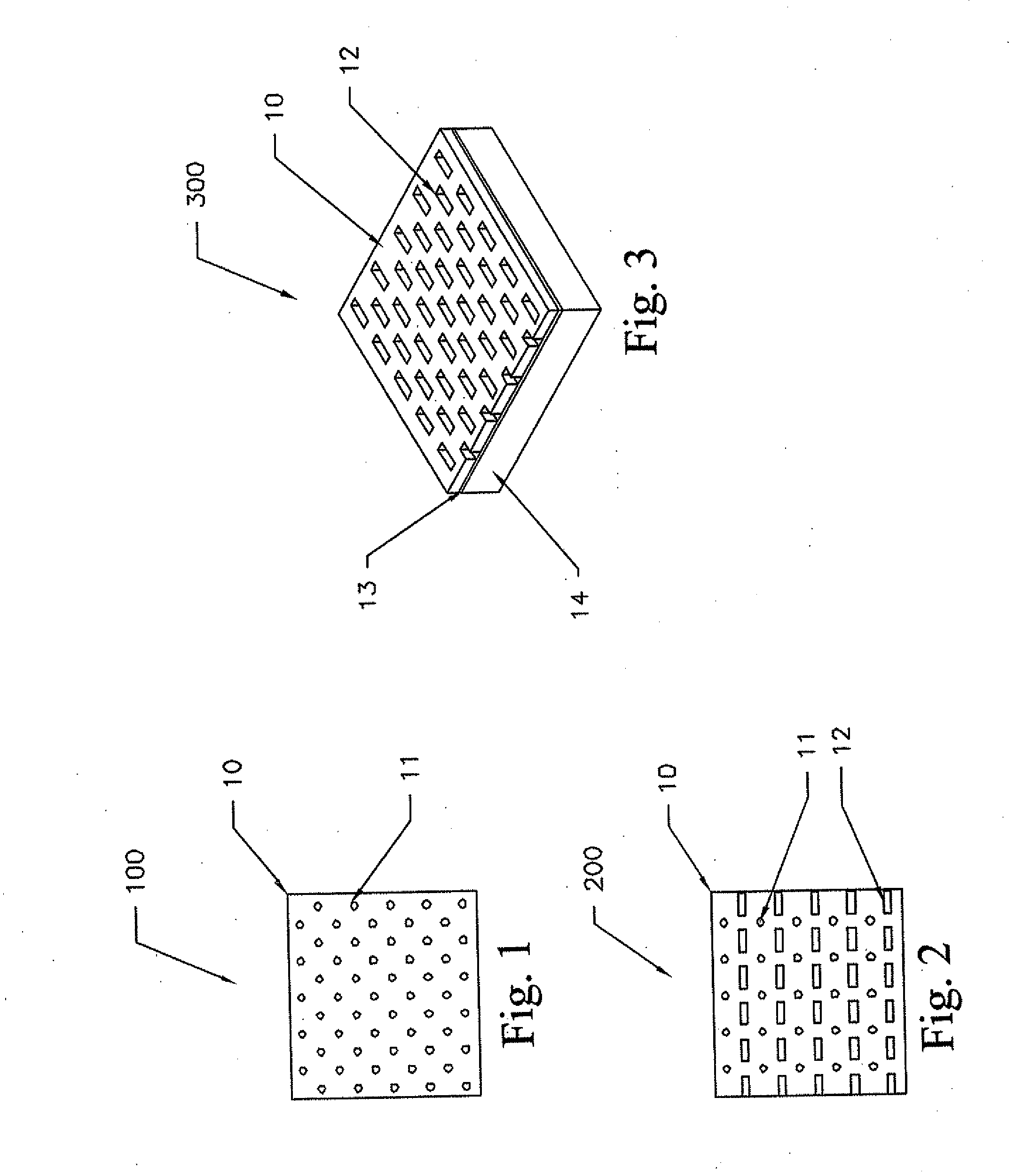
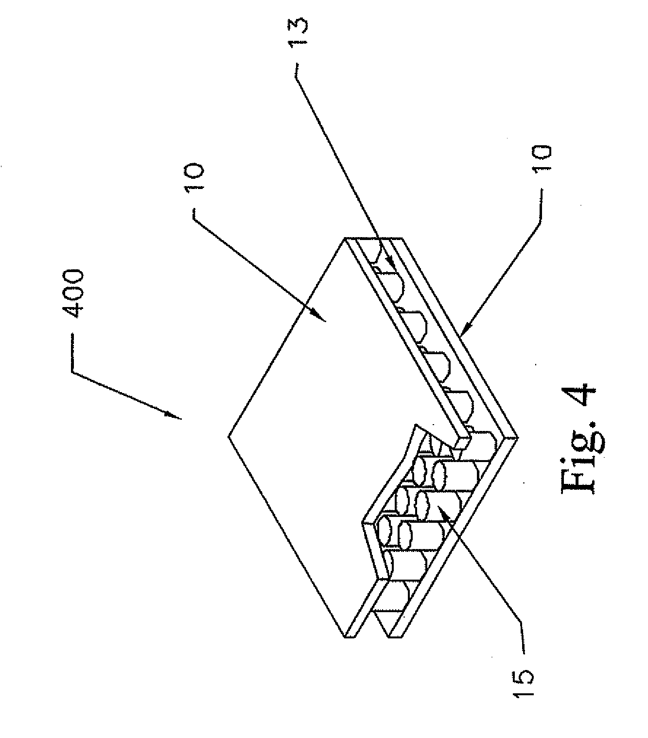

Image

  • Medical device
  • Medical device
  • Medical device

Examples


example 1

[0200]A dressing of the present disclosure was used to treat a 45 year old male suffering from cutaneous manifestation of “shingles”, Herpes zoster virus unilaterally at the tenth thoracic dermatome measuring 2 inches by 3 inches. The patient applied the multilayer wound pad illustrated in FIG. 12 after moistening the pad with tap water. The dressing was held in place with an adhesive layer and backing sheet Within five minutes, the patient reported 25% reduction in pain and within 2 hours nearly 90% reduction in pain. The patient reported that as the dressing dried out the pain returned, but never returned to the level experienced prior to placement of the dressing. When the dressing was re-moistened with water, the pain level was significantly reduced within ten minutes. The dressing was moistened through the moisture regulation layer without removing the dressing from the cutaneous viral outbreaks. The cutaneous lesions healed within 36 hours after application.

example 2

[0201]A three year old female received 80% total body surface area full thickness (third degree burns) burns secondary to a flame injury. She was taken to surgery shortly after admission and all body surface areas were debrided of necrotic tissue. Integral synthetic skin was applied and covered with the wound dressing illustrated in FIG. 12. The dressing was changed every two days leaving the synthetic skin in place. Gradually the synthetic skin was surgically excised and meshed split thickness skin graphs were applied. The wound dressing was applied over the meshed split thickness skin graphs, and was changed every two days until the wounds healed. The dressing was moistened every 12 hours with sterile water throughout the course of healing.

example 3

[0202]Table 1 illustrates the release of silver ions. A four inch by four inch square of an autocatalytic electroless silver plated 5.5 ounce per square yard warp knit fabric was incubated in tryptic soy broth at 37° C. The concentration of silver ions was measured inductively coupled plasma spectroscopy over a twelve day period. FIG. 8 illustrates that the concentration of silver ions increased from less than 10 micrograms / ml the first hour, to over 60 micrograms / ml by day 5.

TABLE 1Time235812Dressing1 Hr2 Hr4 Hr24 HrDayDayDayDayDay4 inch8.513.919.143.151.958.165.464.564.2by 4μg / mlμg / mlμg / mlμg / mlμg / mlμg / mlμg / mlμg / mlμg / mlinch 5.5oz / yd2

[0203]It is well known that between 3 and 25 micrograms / milliliter of ionic silver are required to kill the most common pathologic wound microorganisms. Results indicated that the effective silver ion concentration was attained in about 1 to about 4 hours.


PUM


Abstract

A medical device for example a wound dressing having antibacterial and optionally, antifungal properties, are provided together with methods for making the device. An exemplary dressing includes a layer of silver-containing fabric, (optionally) a layer of absorbent material, and (optionally) a layer of flexible air-permeable and / or water-impermeable material. The dressing can be used for prophylactic and therapeutic care and treatment of skin infections and surface wounds (including surgical incisions), as a packing material, and as a swab for surface cleaning.

Description

CROSS-REFERENCE TO RELATED APPLICATIONS[0001]This application is a continuation-in-part of U.S. patent application Ser. No. 10 / 421,370, filed on Apr. 23, 2004, which is a continuation-in-part application of U.S. patent application Ser. No. 09 / 531,245, filed on Mar. 21, 2000, now U.S. Pat. No. 6,861,570, and is a continuation of PCT / US98 / 19689 filed on Sep. 22, 1998; this application is a continuation-in-part of U.S. patent application Ser. No. 08 / 707,779 filed on Sep. 3, 1996, which is a continuation-in-part application of U.S. patent application Ser. No. 08 / 524,134, filed Sep. 5, 1995, and U.S. patent application Ser. No. 08 / 623,046, filed Mar. 28, 1996; this application is a continuation-in-part of U.S. patent application Ser. No. 10 / 660,209, filed on Sep. 11, 2003, which is a continuation application of U.S. patent application Ser. No. 09 / 531,245, filed on Sep. 22, 1997, now U.S. Pat. No. 6,861,570; this application is a continuation-in-part of U.S. patent application Ser. No. 09...

Claims


Application Information

Patent Timeline
Patent Type & Authority Applications(United States)
IPC IPC(8): A61F13/00
CPCA61F13/00063A61F13/022A61F13/36A61F2013/00119A61F2013/00251A61F2013/00327A61F2013/00936A61F2013/00578A61F2013/00604A61F2013/0074A61F2013/00859A61F2013/0091A61F2013/00553
Inventor FLICK, A. BARTHOLOMEWSILVER, GREGGMILLER, THOMAS
Owner ARGENTUM MEDICAL
Learn More