Method and system for local adaptive detection of microaneurysms in digital fundus images

a technology of microaneurysms and fundus images, applied in the field of medical imaging analysis, can solve the problems of ophthalmologists screening a large number of diabetic patients annually, manual identification of mas in fundus images is time-consuming and subject to inter- and intra-operator variability, and achieves the effect of improving the specificity of ma detection, reducing the workload of ophthalmologists, and improving detection accuracy

Active Publication Date: 2012-01-17
SIEMENS HEALTHCARE GMBH
View PDF10 Cites 16 Cited by
  • Summary
  • Abstract
  • Description
  • Claims
  • Application Information

AI Technical Summary

Benefits of technology

[0018]In order to address the above-described problem, a local adaptive algorithm is proposed for automatic detection of MAs, where multiple subregions of each image are automatically analyzed to adapt to local intensity variations and properties. A priori structural features and pathology, such as region and location information of vessels, optic disk and hard exudates, are further incorporated to improve the detection accuracy. The method effectively improves the specificity of MA detection in digital fundus images, without sacrificing sensitivity. The technique has the potential for use in automatic level-one grading of diabetic retinopathy screening.
[0022]In addition to correcting for shading effect, the step of image enhancing each subregion may also include enhancing local contrast in the subregion, and smoothing to reduce step effects.

Problems solved by technology

Small dotted hemorrhages are often hard to visually differentiate from MAs.
Manual identification of MAs in a fundus image is time-consuming and subjected to inter- and intra-operator variability.
Screening a large number of diabetic patients annually poses a huge workload for ophthalmologists.
Performance of those algorithms is often susceptible to non-uniform illumination and to the locations of MAs in different retinal regions.
Thus, a global processing method often generates a considerable number of false detections.
Some preprocessing techniques, such as shade correction, can ease the severity of inhomogeneous imaging conditions; however, the problems associated with global thresholding still exist.
The region grow, feature extraction and classification steps can remove some false detections, but those steps may also introduce additional errors.
For example, region growing for small objects such as MAs is not very reliable.
The shape feature in MAs detection is essential to classification; however, due to the irregular shape of MAs, the classifier is usually trained to accept shapes varying in a large range, which leads to misclassification.
Those issues exist for MAs detection using fluorescein angiograms, and are likely to be more severe with digital fundus images, where MAs appear to have much weaker contrast with neighboring pixels.
As a result, performance is more sensitive to parameter adjustment, and is less robust.
Several factors, however, may hinder its success in real applications.
The method therefore becomes impractical with digital fundus images, which are normally 1024×1280 pixels.
To the inventors' knowledge, there is currently no such technique available.

Method used


Image

  • Method and system for local adaptive detection of microaneurysms in digital fundus images
  • Method and system for local adaptive detection of microaneurysms in digital fundus images
  • Method and system for local adaptive detection of microaneurysms in digital fundus images

Examples


Embodiment Construction

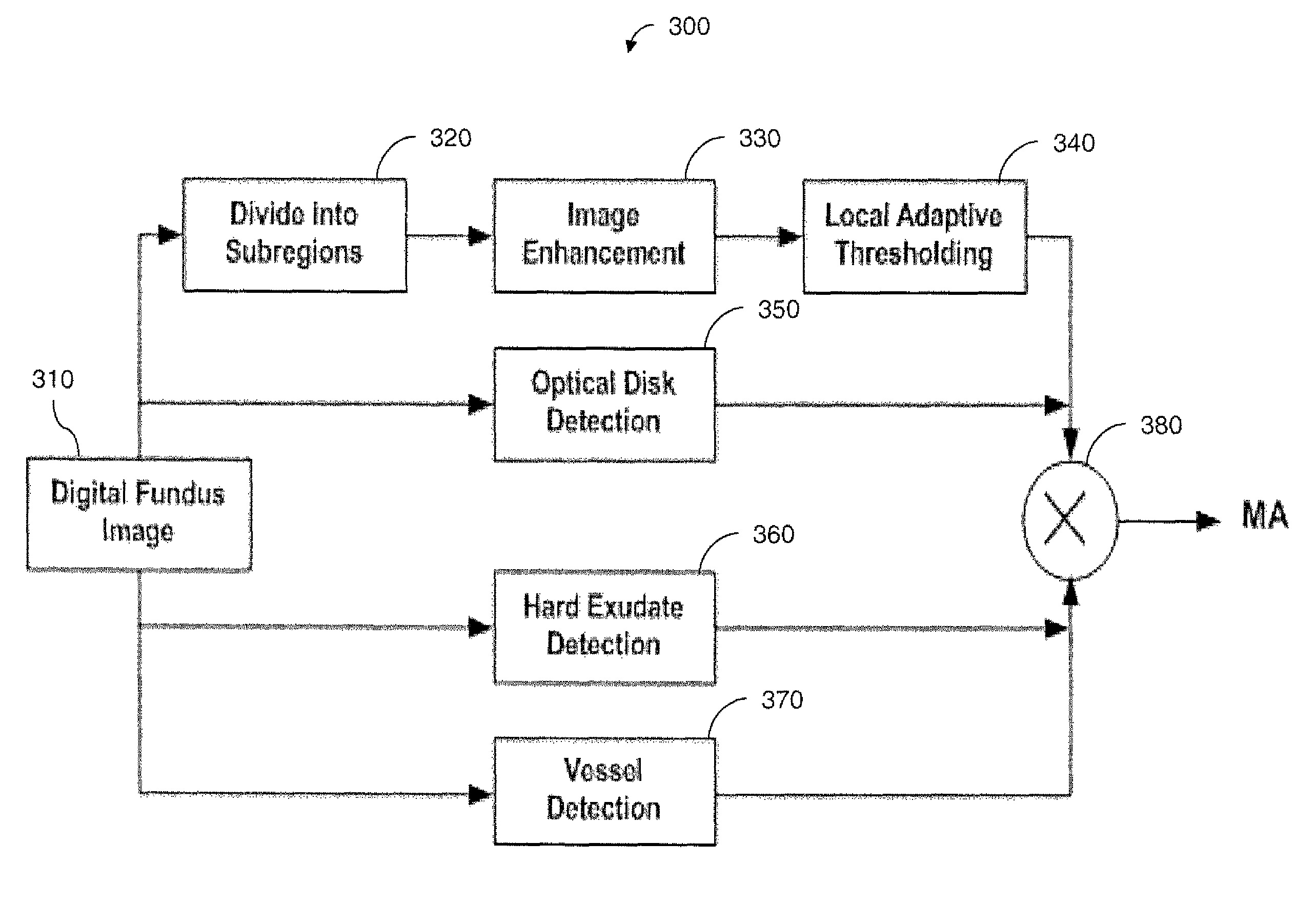
[0034]The inventors have developed a new scheme for robust MAs detection using digital ocular fundus images. The new scheme: (1) takes into account the local properties and variations to improve sensitivity of detection; (2) incorporates a priori knowledge during detection to further reduce false detections (such as, no MAs would appear on blood vessels); and (3) is more robust to parameter selections, and thus to different imaging conditions.

[0035]A flow chart illustrating the inventive scheme 300 is shown in FIG. 3. A fundus image 310 is first automatically subdivided (step 320), and each subregion is then analyzed adaptively (steps 330, 340). Detections of optic disk (step 350), vessel regions (step 370) and hard exudates (step 360) are introduced in parallel to incorporate prior knowledge about locations where MAs would not appear. The a priori information is combined by multiplication (step 380) with the analysis results to yield accurate MAs detection.

[0036]Image Division and ...


PUM


Abstract

A local adaptive method is proposed for automatic detection of microaneurysms in a digital ocular fundus image. Multiple subregions of the image are automatically analyzed and adapted to local intensity variation and properties. A priori region and location information about structural features such as vessels, optic disk and hard exudates are incorporated to further improve the detection accuracy. The method effectively improves the specificity of microaneurysms detection, without sacrificing sensitivity. The method may be used in automatic level-one grading of diabetic retinopathy screening.

Description

CROSS REFERENCE TO RELATED APPLICATIONS[0001]This application claims the benefit of U.S. Provisional Application Ser. No. 60 / 696,242 entitled “A Local Adaptive Algorithm for Microaneurysms Detection in Digital Fundus Images,” filed on Jul. 1, 2005, the contents of which are hereby incorporated by reference herein in their entirety.FIELD OF THE INVENTION[0002]The present invention relates generally to the field of medical imaging analysis, and more particularly, to techniques and systems for automatically identifying microaneurysms (MAs) in digital ocular fundus images.BACKGROUND OF THE INVENTION[0003]Diabetic retinopathy is a widespread eye disease that may cause blindness in diabetic patients. Often patients are not aware of the disease until its late stages, thus annual screening of patients for possible diabetic retinopathy is recommended.[0004]In the screening, microaneurysms (MAs) are one of the earliest visible lesions in diabetic retinopathy, and are therefore an important pa...

Claims


Application Information

Patent Timeline
Patent Type & Authority Patents(United States)
IPC IPC(8): G06K9/00G06K9/42
CPCG06T7/0012A61B3/12G06T2207/30041
Inventor YAN, XIAO-HONG MICHELLEHUANG, KESCHULL, HANS
Owner SIEMENS HEALTHCARE GMBH
Why Eureka
Try Eureka