Anastomosis apparatus and methods

a technology of anastomosis and apparatus, which is applied in the field of anastomosis apparatus and methods, can solve the problems of increased risk of artery trauma caused by ligatures at the clamped site, increased risk of angina and ischemia, and increased discomfort and risks of patients, so as to enhance or form a seal.

Inactive Publication Date: 2012-01-31
MEDTRONIC INC
View PDF665 Cites 28 Cited by
  • Summary
  • Abstract
  • Description
  • Claims
  • Application Information

AI Technical Summary

Benefits of technology

[0019]With this construction, the device can be used to position the first tubular structure (e.g, a graft) in sealing relationship with an opening formed in the second tubular structure prior to passing the fasteners through the first and second tubular structures. The slidably coupled piercing members also improve the procedure for coupling the everted portion of the first tubular structure to the apparatus.
[0020]According to another embodiment, apparatus for anastomosing a first tubular structure to a second tubular structure, having a sidewall with an opening formed therein, comprises a support having a body portion, a plurality of arms extending from the body portion, and a plurality of tissue piercing members each being adapted to pierce a portion of the first and second tubular structures, each of the plurality of arms forming a pathway in which one of the plurality of tissue piercing members is slidably mounted, each of the plurality of arms further having a distal end from which one of the tissue piercing members can be extended and a proximal end, the distal ends are collectively adapted to receive an end portion of the first tubular structure everted thereover. The distal ends can be radially movable so that they can be moved radially outward after introduction in the opening in the second structure to enhance or form a seal between the first tubular structure and the second tubular structure.
[0023]According to a further aspect of the invention, the need for clamping or interrupting fluid flow through the second or target tubular structure can be avoided. In the case where the target tubular structure is a blood carrying vessel (e.g., a patient's aorta), the seal that can be achieved between the first and second tubular structures, which minimizes or eliminates bleeding from the opening in the target tubular structure. Without removing the seal, fasteners can be passed through the spaces or openings between the arms and through the tubular structures to secure the first and second tubular structures together.

Problems solved by technology

The occlusion of the arteries can lead to insufficient blood flow resulting in discomfort and risks of angina and ischemia.
Significant blockage of blood flow in the coronary artery can result in damage to the myocardial tissue or death of the patient.
One problem encountered in either CABG procedure is the need to perform the procedure, while simultaneously maintaining sufficient function of the patient's circulatory system.
Among the drawbacks associated with aortic clamping are an increased chance of trauma to the arteries caused by ligatures at the clamped site and the possible dislodging of plaque within the clamped vessel wall.
Clamping the artery creates a risk of plaque being released into the blood stream.
This release of plaque has the potential of causing a stroke, occlusion of a smaller peripheral vessel, or other vascular trauma.
In a beating heart procedure, full clamping (i.e., cross clamping) of the aorta for graft attachment at the proximal anastomosis is not feasible.
This type of clamping procedure poses the same risks described above with regard to cross clamping, e.g., the risk of release of plaque and resultant cause of a stroke, occlusion of a smaller peripheral vessel, or other vascular trauma.
Drawbacks of using a balloon catheter in creating a seal to divert blood flow include the possibility of disturbing plaque deposits and creating particles in the blood stream, the chance that the balloon catheter may move within the aorta disrupting the seal and resulting in blood loss, and trauma to aortic tissue caused by the pressure needed to create the seal.
However, the Nobles, et al. device requires the withdrawal of the hemostasis device prior to obtaining a tight seal between the graft and vessel.
Therefore, since the area of hemostasis is lost upon the retrieval of the hemostasis device, the artery is open and blood is lost until the sutures are tightened.
Yet another problem related to CABG procedures lies in the procedure of suturing the vessels to create a tight seal.
If one of the tissues is affixed too close to its edge, the suture can tear through the tissue and impair both the tissue and the anastomosis.
Another problem is that, even after proper alignment of the tissue, it is difficult and time consuming to pass the needle through the tissues, form the knot with the suture material, and ensure that the suture material does not become entangled.
These difficulties are exacerbated by the small size of the artery and graft.
Another factor contributing to the difficulty of the CABG procedure is the limited time available to complete the procedure.
If blood flow is not promptly restored, sometimes in as little as 30 minutes, the tissues the artery supplies may experience significant damage or necrosis.
As mentioned above, surgeons are under pressure to reduce the cross-clamp time, yet, an incomplete suture may result in a leak in the tissue approximation between the vessel and graft.
Hence, the suture cannot be hastily performed.
Additionally, the difficulty of suturing a graft to an artery using minimally invasive surgical techniques, where the surgeon uses ports to access the internal organs to perform the procedure, has effectively prevented the safe use of complicated suturing technology in cardiovascular surgical procedures.
In those cases where multiple bypass procedures are performed, the patient will naturally be subject to increased risks as multiple grafts must be sutured to perform the bypass.

Method used


Image

  • Anastomosis apparatus and methods
  • Anastomosis apparatus and methods
  • Anastomosis apparatus and methods

Examples


Embodiment Construction

[0099]Before the present invention is described, it is to be understood that this invention is not limited to particular embodiments or examples described, as such may, of course, vary. Further, when referring to the drawings, like numerals indicate like elements.

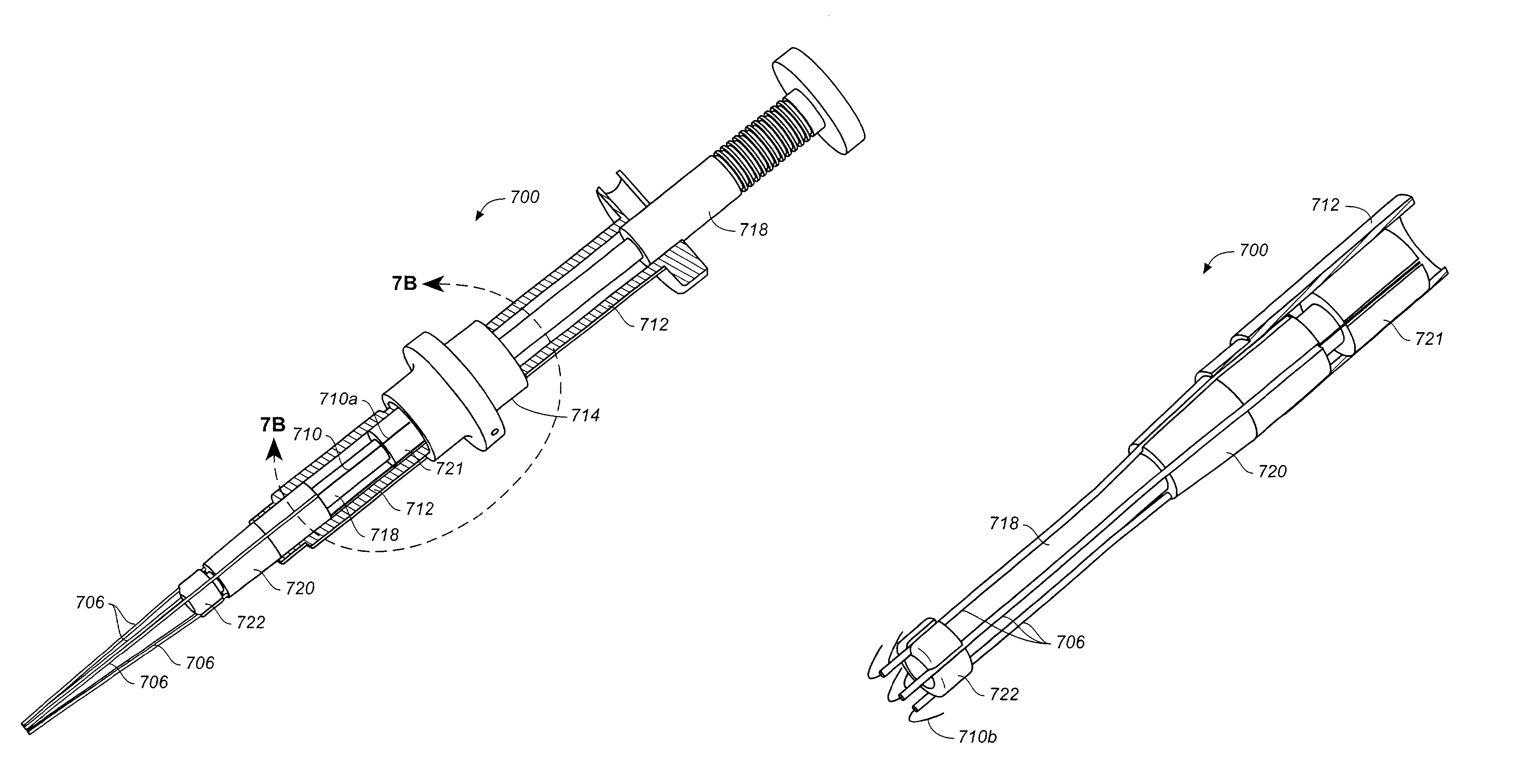
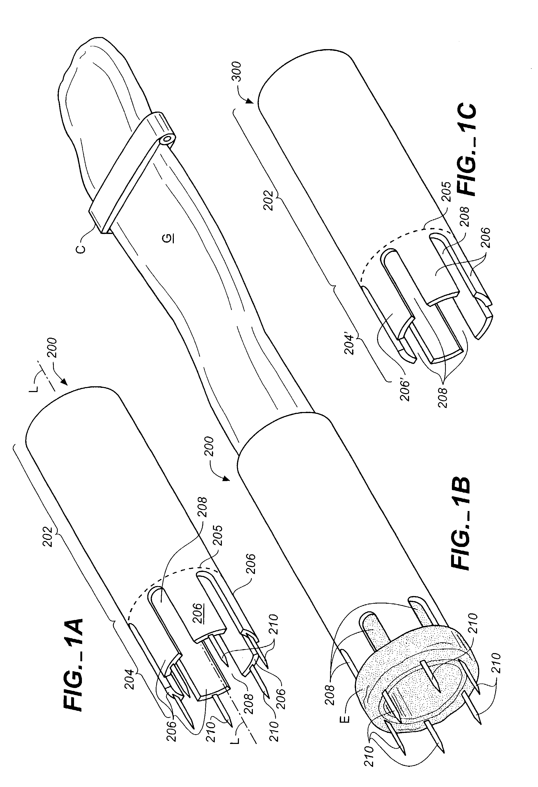
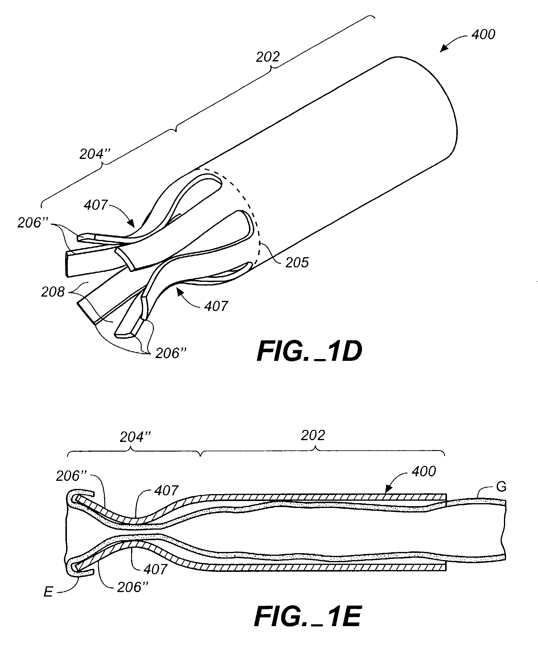
[0100]The devices, systems, and methods described herein can be used to connect or anastomose tubular structures or conduits together. The tubular structures can be vascular or nonvascular structures. The illustrative embodiments will be described in connection with coronary artery bypass grafting procedures during which a vascular conduit or graft structure, such as a vein (e.g., a saphenous vein), artery (e.g., an internal mammary artery), or an artificial conduit or graft structure, is anastomosed to an aorta, the example target structure. It should be understood, however, that the invention can be used in other applications not specifically described herein. For example, the devices also can be used to anastomose intern...


PUM


Abstract

A system for anastomosing a first tubular structure to a second tubular structure having an opening formed therein comprises a support device having a plurality of arms forming spaces therebetween and a plurality of piercing members slidably coupled to the arms. The support device piercing members have a retracted state and an extended state to support the first tubular structure thereon for placement within an opening in the second tubular structure to facilitate anastomosing the tubular structures together. The arms can be configured to urge the first tubular structure against the portion of the second tubular structure surrounding the opening to form a seal therebetween. According to one embodiment, a plurality of discrete fasteners are provided to pass through the support device spaces and the first and second tubular structures. In another embodiment, fasteners are integrated into the support device and releasably coupled thereto for delivery to the anastomosis site.

Description

CROSS-REFERENCE[0001]This application also claims the benefit of U.S. Provisional Application No. 60 / 415,997, filed Oct. 4, 2002, which application is incorporated herein by reference.FIELD OF THE INVENTION[0002]This invention relates to apparatus and methods for joining tubular structures of which at least one is tissue. More particularly, the invention involves anastomosing these structures. One example application is in a proximal anastomosis.BACKGROUND OF THE INVENTION[0003]The occlusion of the arteries can lead to insufficient blood flow resulting in discomfort and risks of angina and ischemia. Significant blockage of blood flow in the coronary artery can result in damage to the myocardial tissue or death of the patient. In most cases, occlusion of the artery results from progressive long term deposits of plaque along the artery wall. While such deposits may be concentrated and occlude the artery at a particular site, the deposits are most certainly present throughout the arter...

Claims


Application Information

Patent Timeline
Patent Type & Authority Patents(United States)
IPC IPC(8): A61B17/08A61B17/04A61B17/06A61B17/064A61B17/11A61B17/115
CPCA61B17/0644A61B17/11A61B17/115A61B17/1152A61B17/0469A61B17/0482A61B2017/0472A61B2017/06009A61B2017/0647A61B2017/1107
Inventor GOLDEN, STEVESCHALLER, LAURENTMAVROIDIS, DEMETRIAINSWORTH, STEPHENWOODSIDE, KATHLEENHO, LIEM
Owner MEDTRONIC INC
Why Eureka
Try Eureka