Homogeneous luminescence bioassay

a bioassay and homogeneous technology, applied in the field of homogeneous luminescence bioassay, can solve the problems of limited further improvement of sensitivity, serious limitations of sensitivity, and significant increase of background signal, and achieve the effect of increasing the fluorescence of said acceptor

Active Publication Date: 2012-05-22
HIDEX
View PDF12 Cites 7 Cited by
  • Summary
  • Abstract
  • Description
  • Claims
  • Application Information

AI Technical Summary

Problems solved by technology

The concentration of the fluorescent conjugate of streptavidin must be carefully adjusted because an excess of it results in a significant increase in the background signal.
The energy transfer was measured from decrease in the fluorescence of the donor, which limited further improvements in sensitivity.
Many compounds and proteins present in biological fluids or serum are intrinsically fluorescent, and the use of conventional fluorophores leads to serious limitations of sensitivity (Wu P and Brand L, Anal Biochem 1994; 218:1-13).
Another major problem with homogeneous fluorescence techniques is the inner filter effect and the variability of the optical properties of a sample.
Sample dilution has been used to correct this drawback, but always at the expense of analytical sensitivity.
However, still only a small part of the lanthanide chelates inside the entire particulate can participate in an energy transfer to a single acceptor and thus the entire fluorescence of a particulate label cannot be quenched by a single acceptor moiety.
The same problem is also encountered when lanthanide chelates are incorporated in a solid phase.
In most of the conventional homogeneous fluorescence assay technologies, the performance has still severe limitations: the sensitivity is limited by interferences from matrix components and optical properties of matrices, e.g. urine, saliva, serum, plasma or whole blood, to fluorescence yield and level of background, and by the attainable degree of fluorescence modulation, e.g. quenching, enhancement, energy transfer or polarization (Hemmilä I, Clin Chem 1985; 31: 359-370).

Method used


Image

  • Homogeneous luminescence bioassay
  • Homogeneous luminescence bioassay
  • Homogeneous luminescence bioassay

Examples


example 1

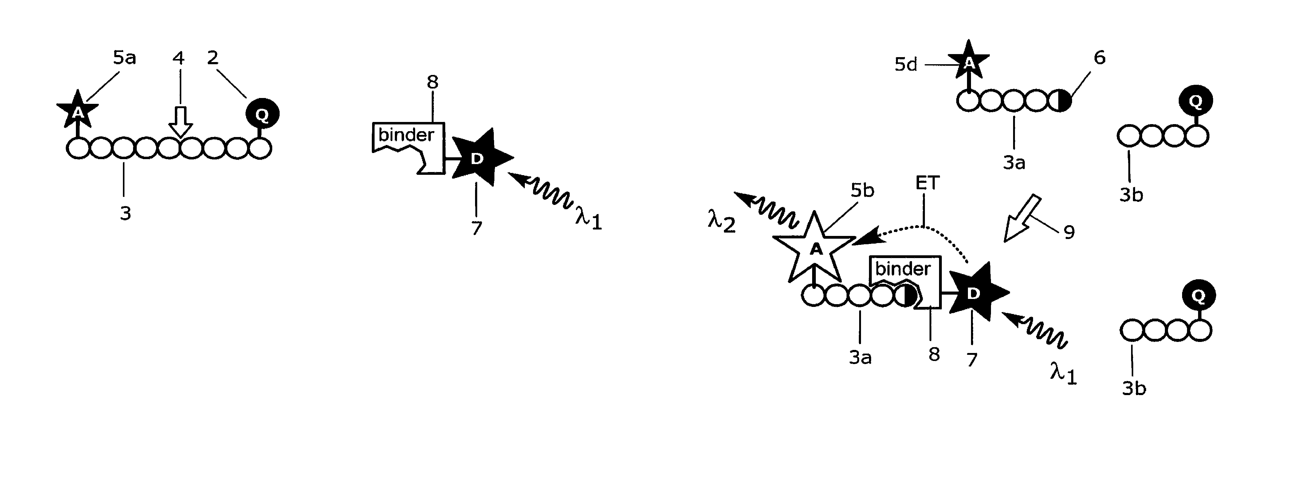
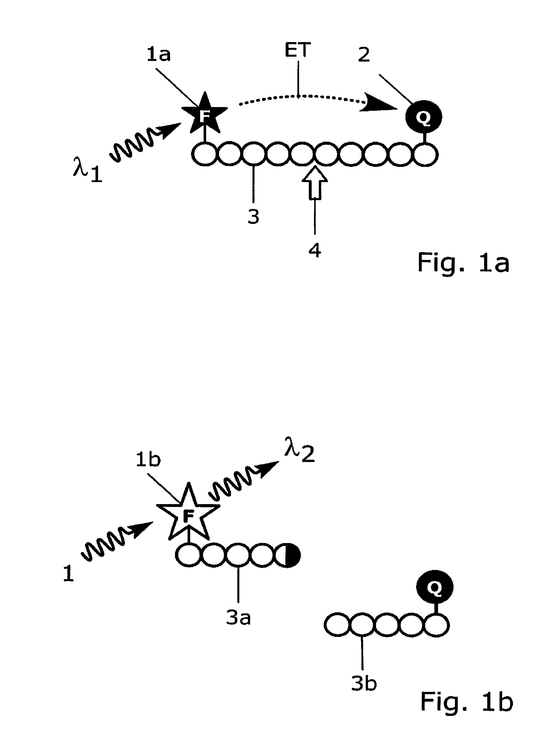
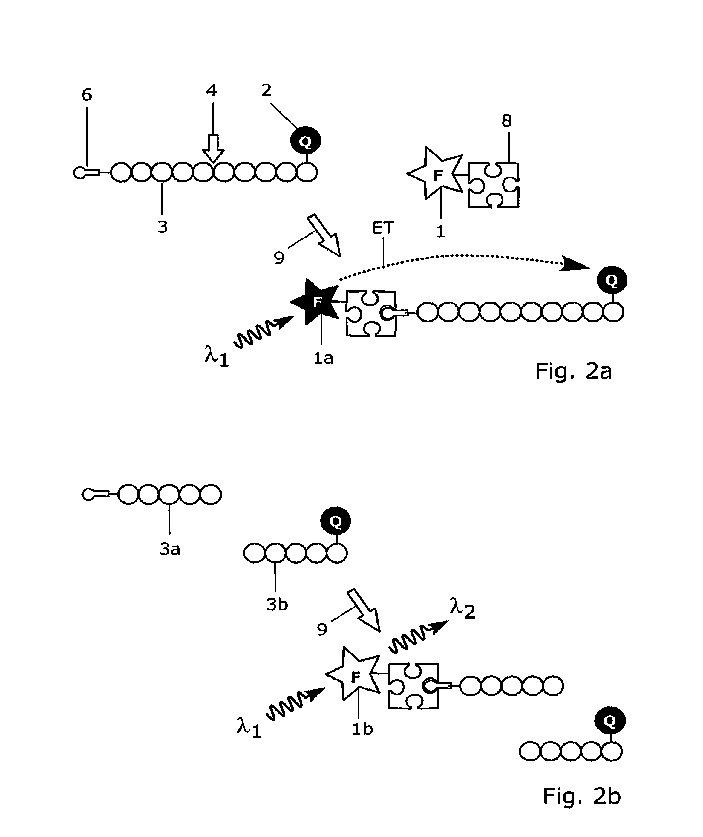
[0179]Antibody 11H3 (0197-100 / bA4N-11H3, NanoTools Antikorpertechnik, Germany; indicated with label 8 in FIGS. 9a and 9b) that specifically recognizes the free N-terminal end of the amyloid peptide βA4 (indicated with label 6 in FIG. 9b) is conjugated to Europium(III)-chelate dyed Fluoromax nanoparticles (indicated with label 7 in FIGS. 9a and 9b) 92 nm in diameter, that is obtained from Seradyn Inc (Indianapolis, Ind.). The conjugation is done as described by Suokka, et al. Anal Chem 2001; 73:2254-60. Nanoparticles are prewashed with 10 mmol / l phosphate buffer, pH 7.0, on a Nanosep microporous centrifugal filter (300 kDa cut off; Pall Filtron, Northborough, Mass.). Phosphate buffer is added to the particles, and the solution is sonicated with a tip sonicator (Labsonic U; B. Braun, Leverkusen, Germany) at 80 W for 5 s. Carboxyl groups on the surface of nanoparticles are activated with 10 mmol / l N-(3-dimethylaminopropyl)-N9-ethylcarbodiimide and N-hydroxysulfosuccinimide (Fluka, Buch...

example 2

[0186]The anti-amyloid peptide 134A antibody 11H3 (indicated with label 8 in FIGS. 9a and 9b) is labelled with fluorescent europium chelate labelling reagent {2,2′,2″, 2′″-{[2-(4-isothiocyanatophenyl)ethylimino]bis(methylene)bis{4-{[4-(α-ga-lactopyranoxy)phenyl]ethynyl}pyridine-6,2-diyl}bis(methylenenitrilo)}tetrakis(ace-tato)}europium(III) (von Lode P et al., Anal Chem 2003; 75: 3193-201; indicated with label 7 in FIGS. 9a and 9b). The labelling of antibody 11H3 is performed in 50 mmol / l sodium carbonate buffer, pH 9.8, using 50-fold molar excesses of the chelate labelling reagent. The reaction is carried out overnight at room temperature and the excess free labelling reagent is removed on NAP-5 and NAP-10 (Amersham Biosciences) chromatography columns using Tris-saline-azide (6.1 g / l Tris, 9.0 g / l NaCl, and 0.5 g / l NaN3), pH 7.75, as elution buffer. The fractions containing the antibody are collected and the europium concentrations are measured against a europium calibrator using D...

example 3

[0188]An assay according to the present invention for the detection of calcium binding related conformational changes in calmodulin was developed (FIGS. 8a and 8b). For the assay, a dual-labelled human calmodulin (indicated with labels 3 and 3c in FIG. 8a) is produced by labelling the amino terminal and carboxy terminal domains of calmodulin with QSY35 dye acting as a quencher compound (indicated with label 2 in FIGS. 8a and 8b) and with Cy5 dye acting as an acceptor group (indicated with label 5a in FIGS. 8a and 8b), respectively. The labelling strategy is adapted from Lang et al., Anal. Biochem. 2005, 342: 271-279, and involves targeting of the first dye to free thiol group of a cysteine engineered to the surface of the protein and the other dye to an engineered selenomethionine residue. For labelling, engineered calmodulin containing mutations threonine 34 to methionine and threonine 110 to cysteine is constructed by site-directed mutagenesis as described by Lang et al., (Anal. B...


PUM

PropertyMeasurementUnit
diameteraaaaaaaaaa
diameteraaaaaaaaaa
diameteraaaaaaaaaa

Abstract

A homogenous bioassay including i) a first group containing a short lifetime fluorescent acceptor, and ii) a second group containing a quencher, with the first and second groups linked by at least a first linkage. The bioassay measures the acceptor's fluorescence increase resulting from cleavage of the first linkage and also includes iii) a third group containing a donor for energy transfer to the acceptor, where the donor is an up-conversion fluorescent compound, a long-lifetime fluorescent compound or an electrogenerated luminescent compound. A conformational or terminal epitope is created on the first group through linkage cleavage, and the third group includes a binder with affinity for this epitope. The acceptor's fluorescence is caused by exciting the donor. Also disclosed are bioassay kits for this method.

Description

[0001]This application is a National Stage of International Application PCT / FI2008 / 050088, filed Feb. 26, 2008, which claims benefit under 35 U.S.C. §119 of U.S. provisional application 60 / 903,514, filed Feb. 27, 2007, and Finnish patent application 20070163, filed Feb. 27, 2007.FIELD OF THE INVENTION[0002]This invention relates to measurement of biological activity or its modulation or analyte concentration using a luminescence energy transfer based homogeneous bioassay.BACKGROUND OF THE INVENTION[0003]The publications and other materials used herein to illuminate the background of the invention, and in particular, cases to provide additional details respecting the practice, are incorporated by reference.[0004]A number of assays based on bioaffinity binding reactions or enzymatically catalyzed reactions have been developed to analyze biologically important compounds or their activity or their biological effect or its modulation from various biological samples (such as serum, blood,...

Claims


Application Information

Patent Timeline
Patent Type & Authority Patents(United States)
IPC IPC(8): C12Q1/68
CPCG01N33/542
Inventor SOUKKA, TEROLAMMINMAKI, URPO
Owner HIDEX
Why Eureka
Try Eureka