
Myelination of congenitally dysmyelinated forebrains using oligodendrocyte progenitor cells

a technology of oligodendrocyte progenitor cells and myelination, which is applied in the field of myelination of congenitally dysmyelinated forebrains, can solve the problems of patchy remyelination, low efficiency, and inability to properly develop or repair myelin, and achieves neither undesired phenotypes nor parenchymal aggregates, and widespread and high-efficiency my

Inactive Publication Date: 2012-06-26
CORNELL RES FOUNDATION INC
View PDF10 Cites 22 Cited by
  • Summary
  • Abstract
  • Description
  • Claims
  • Application Information

AI Technical Summary

Problems solved by technology

In the pediatric leukodystrophies, in particular, compact myelin either fails to properly develop, or is injured in the setting of toxic storage abnormalities.
However, these unfractionated implants yielded only patchy remyelination and would have permitted the co-generation of other, potentially undesired phenotypes.
Neurosci Res 34:1-13 (1993)), though with low efficiency, likely due to predominantly astrocytic differentiation by the grafted cells.

Method used


Image

  • Myelination of congenitally dysmyelinated forebrains using oligodendrocyte progenitor cells
  • Myelination of congenitally dysmyelinated forebrains using oligodendrocyte progenitor cells
  • Myelination of congenitally dysmyelinated forebrains using oligodendrocyte progenitor cells

Examples


example 1

Cells

[0052]Cells Tissue from late gestational age human fetuses (21 to 23 weeks) were obtained at abortion. The forebrain ventricular / subventricular zones were rapidly dissected free of the remaining brain parenchyma, and the samples chilled on ice. The minced samples were then dissociated using papain / DNAse as previously described (Keyoung et al., “Specific Identification, Selection and Extraction of Neural Stem Cells from the Fetal Human Brain,”Nature Biotechnology 19:843-850 (2001), which is hereby incorporated by reference in its entirety), always within 3 hours of extraction. The dissociates were then maintained overnight in minimal culture media of DMEM / F12 / N1 with 20 ng / ml FGF.

example 2

Sorting

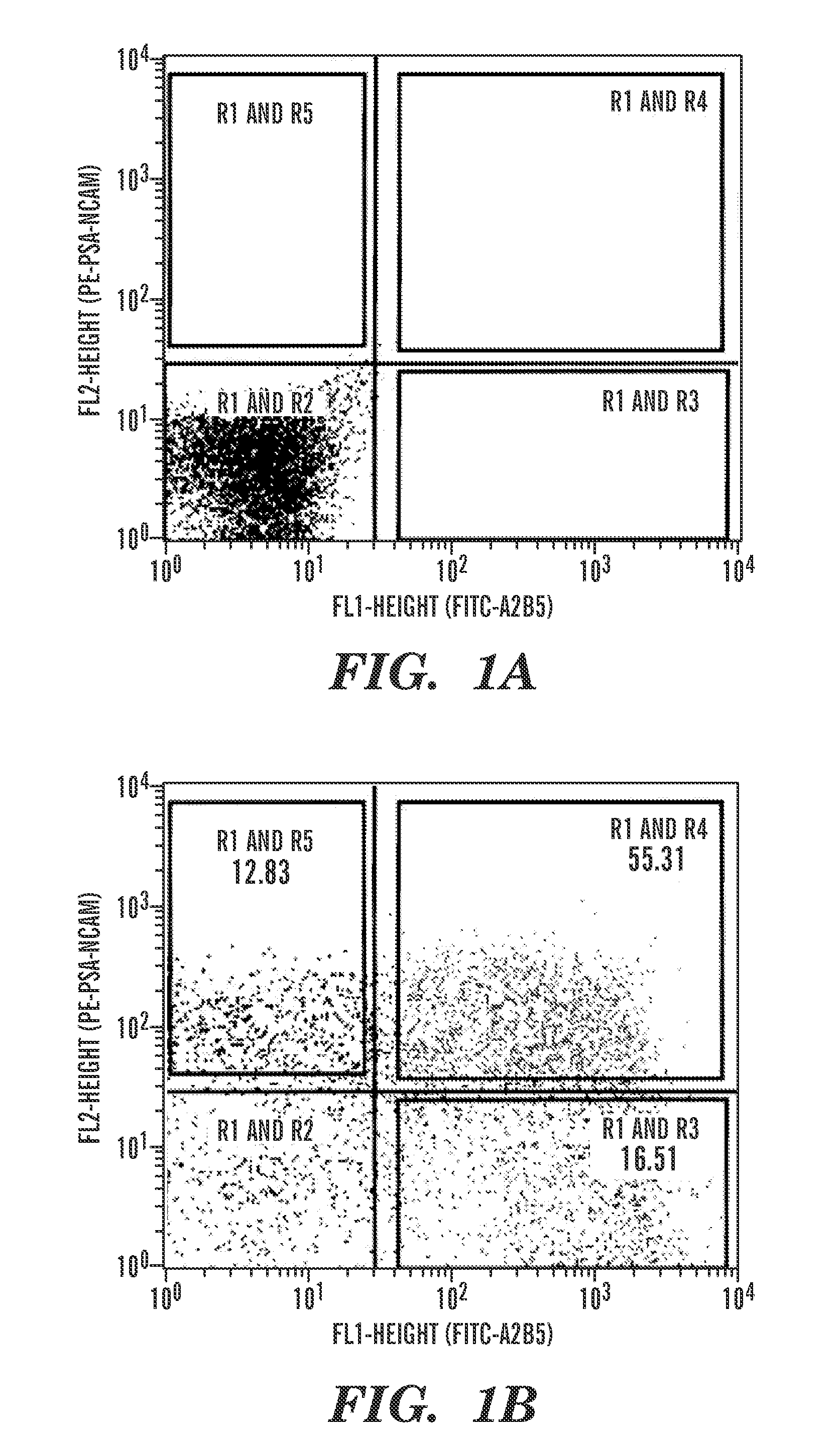
[0053]The day after dissociation, the cells were incubated 1:1 with MAb A2B5 supernatant (clone 105; ATCC, Manassas, Va.), for 30 minutes, then washed and labeled with microbead-tagged rat anti-mouse IgM (Miltenyi Biotech). All incubations were done at 4° C. on a rocker. In some instances, 2-channel fluorescence-activated cell sorting was done to define the proportions and phenotypic homogeneity of A2B5 and PSA-NCAM-defined subpopulations, using a FACSVantage SE / Turbo, according to previously described methods (Keyoung et al., “Specific Identification, Selection and Extraction of Neural Stem Cells from the Fetal Human Brain,”Nature Biotechnology 19:843-850 (2001) and Roy et al., “In Vitro Neurogenesis by Progenitor Cells Isolated from the Adult Human Hippocampus,”Nat Med 6:271-277 (2000), which are hereby incorporated by reference in their entirety). More typically, and for all preparative sorts for transplant purposes, magnetic separation of A2B5+ cells (MACS; Miltenyi) was ...

example 3

Transplantation and Tagging

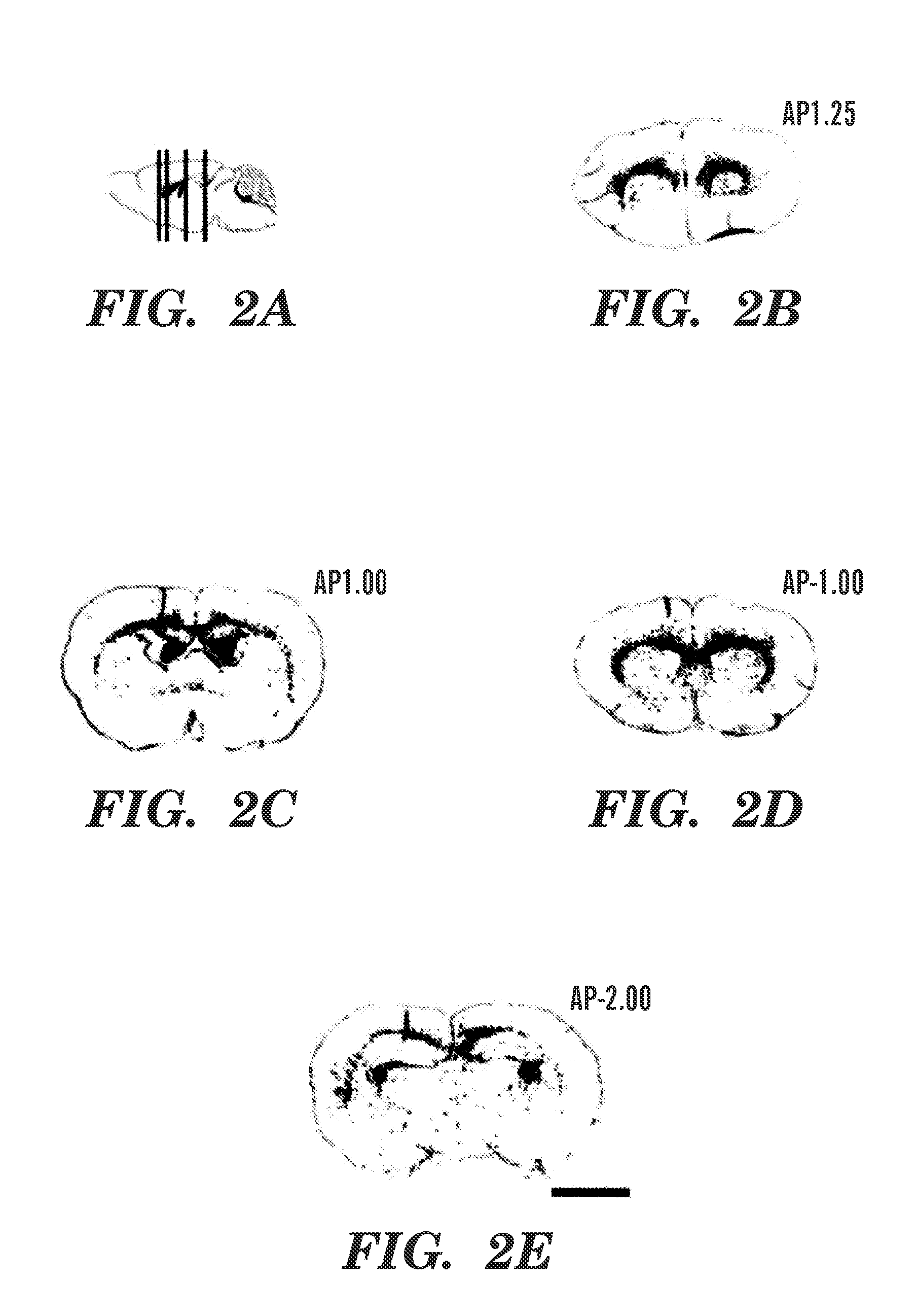
[0054]Homozygous shiverers were bred in a colony. Within a day of birth, the pups were cryoanesthetized for cell delivery. The donor cells were then implanted using a pulled glass pipette inserted through the skull, into either the corpus callosum, the internal capsule, or the lateral ventricle. The pups were then returned to their mother, and later killed after 4, 8, 12, or 16 weeks. For some experiments, recipient mice were injected for 2 days before sacrifice with BrdU (100 μg / g, as a 1.5 mg / 100 μl solution), q12 hrs for 2 consecutive days.


PUM

PropertyMeasurementUnit
widthaaaaaaaaaa
lengthaaaaaaaaaa
volumeaaaaaaaaaa

Abstract

One form of the present invention is directed to a method of remyelinating demyelinated axons by treating the demyelinated axons with oligodendrocyte progenitor cells under conditions which permit remyelination of the axons. Another aspect of the present invention relates to a method of treating a subject having a condition mediated by a loss of myelin or a loss of oligodendrocytes by administering to the subject oligodendrocyte progenitor cells under conditions effective to treat the condition mediated by a loss of myelin or a loss of oligodendrocytes. A further aspect of the present invention relates to an in vitro method of identifying and separating oligodendrocyte progenitor cells from a mixed population containing other mammalian brain or spinal cord cell types. This further aspect of the present invention involves removing neurons and neuronal progenitor cells from the mixed population to produce a treated mixed population. Oligodendrocyte progenitor cells are then separated from the treated mixed population to form an enriched population of oligodendrocyte progenitor cells.

Description

[0001]This application is a continuation of U.S. patent application Ser. No. 10 / 368,810, filed Feb. 14, 2003, which claims the benefit of U.S. Provisional Patent Application Ser. No. 60 / 358,006, filed Feb. 15, 2002, which are hereby incorporated by reference in their entirety.[0002]The subject matter of this invention was made with government support under grant number NINDS R01NS39559 awarded by National Institutes of Health. The government has certain rights in this invention.FIELD OF THE INVENTION[0003]The present invention is directed to the myelination of congenitally dysmyelinated forebrains using oligodendrocyte progenitor cells and to a method of treating a subject having a condition mediated by a loss of myelin or a loss of oligodendrocytes. Also disclosed is a method for the identification and separation of oligodendrocyte progenitor cells from a mixed population containing other mammalian brain or spinal cord cell types.BACKGROUND OF THE INVENTION[0004]A broad range of di...

Claims


Application Information

Patent Timeline
Patent Type & Authority Patents(United States)
IPC IPC(8): A01N63/00A01N65/00A61K31/7052C12N15/09A61K35/12A61K35/76A61P25/00A61P31/04A61P43/00C12N5/02C12N5/079C12N5/0797C12N5/10C12Q1/04
CPCC12N5/0623C12N5/0622A61K35/12C12N2510/00A61P25/00A61P27/02A61P29/00A61P31/04A61P43/00A61P7/04Y02A50/30C12N5/0618A61K48/00A61K35/30C12N5/0619G01N33/56966G01N33/5005
Inventor GOLDMAN, STEVEN A.ROY, NEETA SINGHWINDREM, MARTHA
Owner CORNELL RES FOUNDATION INC
Learn More