Bioactive stents for type II diabetics and methods for use thereof

a bioactive stent and diabetic technology, applied in the field of biodegradable polymer coated implantable stents, can solve the problems of exacerbated injury, increased thrombosis of damaged arterial surfaces within the vascular system, and precipitated new smooth muscle cell proliferation and neointimal growth

Inactive Publication Date: 2006-01-19
MEDIVAS LLC
View PDF13 Cites 97 Cited by
  • Summary
  • Abstract
  • Description
  • Claims
  • Application Information

AI Technical Summary

Problems solved by technology

However, damaged arterial surfaces within the vascular system are highly susceptible to thrombus formation.
However, these techniques exacerbate the injury, precipitating new smooth muscle cell proliferation and neointimal growth.
The effectiveness of this procedure is limited in some patients because the treatment itself damages the vessel, thereby inducing proliferation of smooth muscle cells and reocclusion or restenosis of the vessel.
Damage to the endothelial and medial layers of a blood vessel, such as often occurs in the course of balloon angioplasty and stent procedures, has been found to stimulate neointimal proliferation, leading to restenosis of atherosclerotic vessels.
However, tissue surrounding a porous stent tends to infiltrate the pores.
In certain applications, pores that promote tissue ingrowth are considered to be counterproductive because the growth of neointima can occlude the artery, or other body lumen, into which the stent is being placed.
An unfortunate consequence of this procedure is the nearly total destruction of the endothelial layer by expansion of the angioplasty balloon and precipitation of a foreign body inflammatory response to the stent.
Since mechanical intervention has destroyed the natural blood / artery barrier, in a significant number of patients the result is a local uncontrolled proliferative response by smooth muscle cells leading to restenosis.

Method used


Image

  • Bioactive stents for type II diabetics and methods for use thereof
  • Bioactive stents for type II diabetics and methods for use thereof
  • Bioactive stents for type II diabetics and methods for use thereof

Examples


example 1

[0205] Amide Bond Formation—This example illustrates the coupling of a carboxyl group of a polymer with an amino functional group of the bioactive agent, or equally, the coupling of a carboxyl group of the bioactive agent with an amino functional group of a polymer.

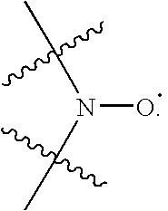
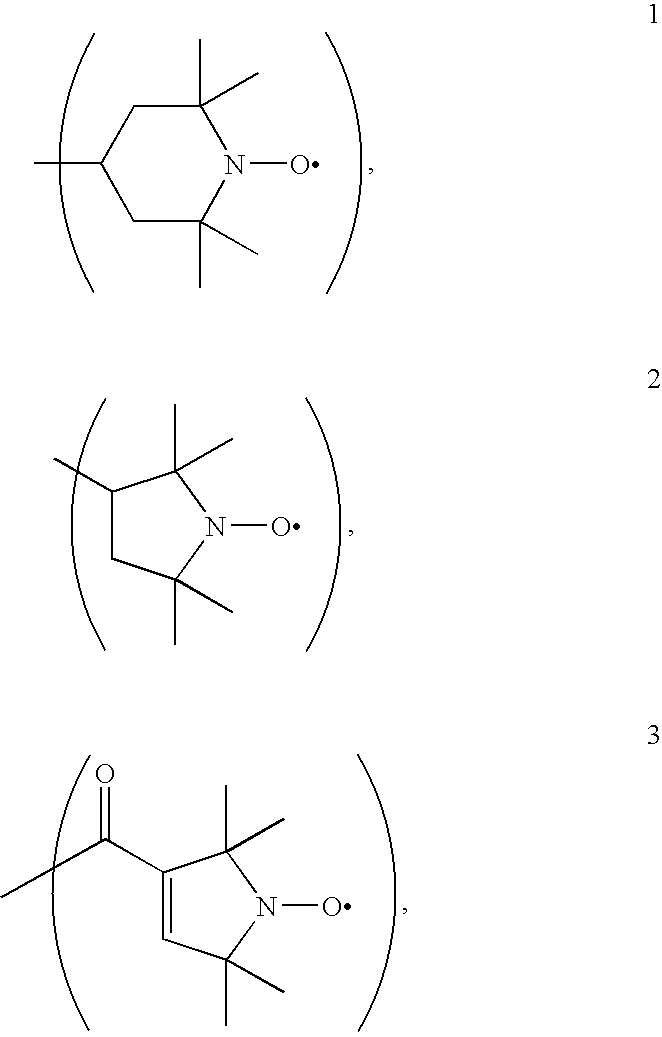
[0206] Coupling Through Pre-Formed Active Esters; Carbodiimide Mediated Couplings—Conjugation of 4-Amino-Tempo to Polymer The free carboxylic acid form of the PEA polymer is converted first to its active succinimidyl ester (PEA-OSu) or benzotriazolyl ester (PEA-OBt). This conversion can be achieved by reacting dried PEA-H polymer with N-Hydroxysuccinimide (NHS) or 1-Hydroxybenzotriazole (HOBt) and a suitable dehydrating agent, such as dicyclohexylcarbodiimide (DCC), in anhydrous CH2Cl2 at room temperature for 16 hrs. After filtering away the precipitated dicyclohexylurea (DCU), the PEA-OSu product may be isolated by precipitation, or used without further purification, in which case the PEA-OSu solution is transferred to ...

example 2

[0208] Ester Bond Formation—This example illustrates coupling of a carboxyl group of a polymer with a hydroxyl functional group of the bioactive agent, or equally, coupling of a carboxyl group of the bioactive agent with a hydroxyl functional group of a polymer.

[0209] Carbodiimide Mediated Esterification For the conjugation, a sample of the carboxyl-group-containing polymer was dissolved in DCM. To this slightly viscous solution was added a solution of the hydroxyl-containing-drug / biologic and DMAP in DCM. The flask was then placed in an ice bath and cooled to 0° C. Next, a solution of 1,3-diisopropylcarbodiimide (DIPC) in DCM was added, the ice bath removed, and the reaction warmed to room temperature. The conjugation reaction was stirred at room temperature for 16 hours during which time TLC was periodically performed to monitor consumption of the hydroxyl functional group of the bioactive agent. After the allotted time, the reaction mixture was precipitated, and the polymer-bioa...

example 3

[0210] PEC Isolations To establish the protocol for isolating the progenitor endothelial cells (PECs) from peripheral blood, blood from healthy, normal donors was used. A literature review generated multiple PEC isolation protocols (J.C.I. (2000) 105:71-77 ; Circ. (2003) 107:143-149; Circ. (2003) 107:1164-1169; Plast. Reconstruc. Surgr. (2004) 113:284; and Am. J. Physiol. Heart Circ. Physiol. (2004) 286:H1985-H1993). Surprisingly, however, preliminary attempts required modification of the known protocols to ensure successful isolations. The flow chart in FIG. 2 presents a modified protocol followed in isolation of PECs.

[0211] From a trial PEC isolation, it was determined that cells would attach and grow better on fibronectin-coated plates than on gelatin-coated plates. Cells were isolated from ˜120 milliliters of peripheral blood and then single aliquots of cells were plated in Endothelial Basal Medium and 5% FBS (Cambrex). The media was changed every 4-5 days. The total cell numbe...


PUM

PropertyMeasurementUnit
temperatureaaaaaaaaaa
reaction timeaaaaaaaaaa

Abstract

The present invention is based on the discovery that a vascular stent or other implantable medical device can be coated with a biodegradable biocompatible polymer to which is attached a bioligand that specifically captures progenitors of endothelial cells (PECs) from the circulating blood to promote endogenous formation of healthy endothelium in Type II diabetics. In one embodiment, the bioligand is a peptide that specifically binds to an integrin receptor on PECs. The invention also provides methods for using such vascular stents and other implantable devices to promote vascular healing in Type II diabetics, for example following mechanical intervention.

Description

RELATED APPLICATIONS [0001] This application is a Continuation-in-Part application of U.S. patent application Ser. No. 11 / 098,891, filed Apr. 4, 2005 and relies for priority under 35 U.S.C. § 119(e) upon U.S. Provisional Application Ser. No. 60 / 559,937, filed Apr. 5, 2004.FIELD OF THE INVENTION [0002] The invention relates generally to implantable medical devices, and in particular to biodegradable polymer coated implantable stents that promote vascular healing in diabetics. BACKGROUND INFORMATION [0003] The normal endothelium, which lines blood vessels, is uniquely and completely compatible with blood. Endothelial cells initiate metabolic processes, like the secretion of prostacyclin and endothelium-derived relaxing factor (EDRF), which actively discourage platelet deposition and thrombus formation in vessel walls. However, damaged arterial surfaces within the vascular system are highly susceptible to thrombus formation. Abnormal platelet deposition, resulting in thrombosis, is mor...

Claims


Application Information

Patent Timeline
Patent Type & Authority Applications(United States)
IPC IPC(8): A61F2/00A61F2/82A61K39/395C07K16/18C07K16/28
CPCA61F2/82A61F2250/0068A61L27/34A61L27/56A61L31/10C07K16/18C07K16/28C08L89/00
Inventor CARPENTER, KENNETHTURNELL, WILLIAMDEFIFE, KRISTINGRAKO, KATHRYN
Owner MEDIVAS LLC
Why Eureka
Try Eureka