System and method for assessing coupling between an electrode and tissue

a tissue and electrode technology, applied in the field of system and method for assessing the degree of coupling between an electrode and a tissue, can solve the problems of ineffective lesions, affecting the effect of electrodes, and affecting the effectiveness of lesions, etc., and achieves the effect of improving the degree of coupling, facilitating interpretation and correlation, and conducting more efficiently

Active Publication Date: 2013-05-28
ST JUDE MEDICAL ATRIAL FIBRILLATION DIV
View PDF94 Cites 133 Cited by
  • Summary
  • Abstract
  • Description
  • Claims
  • Application Information

AI Technical Summary

Benefits of technology

[0016]A system for assessing a degree of coupling between an electrode and a tissue in a body in accordance with one embodiment of the present invention includes an electronic control unit configured to acquire values for first and second components of a complex impedance between the electrode and the tissue. The first and second components may comprise, for example, a resistance between the electrode and the tissue and a reactance between the electrode and the tissue. Alternatively, the first and second components may comprise an impedance magnitude between the electrode and the tissue and an impedance phase angle. The electronic control unit is further configured to calculate a coupling index responsive to the values. The coupling index is indicative of a degree of coupling between the electrode and the tissue. In accordance with one aspect of the invention, the coupling index may be displayed to a clinician on a display device in a variety of ways to provide to permit easy interpretation and correlation of the measured impedance to coupling between the electrode and tissue.
[0019]The above-described system, article of manufacture and method are advantageous because they provide a true measure of impedance at the interface of the electrode and the target tissue and, therefore, provide a better assessment of coupling between the electrode and the tissue. Further, the system, article of manufacture and method provide an indicator of coupling (i.e., the coupling index) in a format that allows easy interpretation and correlation to tissue contact by the clinician.
[0020]The coupling index can be used in a wide variety of diagnostic and therapeutic devices. In ablation catheters, for example, procedures can be conducted more efficiently and with higher success rates and fewer complications because the coupling index provides an indication of the energy delivered to the target tissue and the proximity of the catheter and electrode to the target tissue and may also enable a clinician to identify the orientation or angle of the ablation electrode relative to the tissue. The index also can act as a proximity sensor indicating the distance between a diagnostic or therapeutic device and a target tissue. For example, in a transseptal access sheath, the coupling index provides an indication that the sheath is approaching (and potentially slipping away from) the septum. In a mapping catheter, the coupling index may improve geometric modeling of the tissue surface by, for example, identifying the most relevant electrode readings and / or allowing multiple electrodes to simultaneously obtain relevant measurements.

Problems solved by technology

Arrhythmia can create a variety of dangerous conditions including irregular heart rates, loss of synchronous atrioventricular contractions and stasis of blood flow which can lead to a variety of ailments and even death.
If the electrode is too far from the tissue or has insufficient contact with the tissue, the lesions created may not be effective.
On the other hand, if the catheter tip containing the electrode contacts the tissue with excessive force, the catheter tip may perforate or otherwise damage the tissue (e.g., by overheating).
Each of these methods has disadvantages, however.
The determination is particularly difficult when using catheters that are relatively long (such as those used to enter the left atria of the heart).
Because fluoroscopic images are two-dimensional projections and blood and myocardium attenuate x-rays similarly, it is difficult to quantify the degree of contact and to detect when the catheter tip is not in contact with the tissue.
Intracardiac echo is time consuming and it is also difficult to align the echo beam with the ablation catheter.
Further, intracardiac echo does not always permit the clinician to confidently assess the degree of contact and can generate unacceptable levels of false positives and false negatives in assessing whether the electrode is in contact with tissue.
Atrial electrograms do not always correlate well to tissue contact and are also prone to false negatives and positives.
Pacing thresholds also do not always correlate well with tissue contact and pacing thresholds are time-consuming and also prone to false positives and false negatives because tissue excitability may vary in hearts with arrhythmia.
Evaluating lesion size at necropsy is seldom available in human subjects, provides limited information (few data points) and, further, it is often difficult to evaluate the depth and volume of lesions in the left and right atria.
Finally, temperature measurements provide limited information (few data points) and are difficult to evaluate in the case of irrigated catheters.
Contact force, however, does not directly measure how well electrical energy is coupled between the catheter electrode and tissue.
This measurement, however, does not correlate well with the more localized contact between the catheter electrode and tissue because it measures the impedance provided not just by the local target tissue, but the entire impedance from the electrode to a cutaneous return electrode through various body tissues and fluids.
Furthermore, generator reported impedance is usually infrequently obtained and at low resolution (about 1Ω).
It is also not readily available to the clinician in a format that allows easy interpretation and correlation to tissue contact.

Method used


Image

  • System and method for assessing coupling between an electrode and tissue
  • System and method for assessing coupling between an electrode and tissue
  • System and method for assessing coupling between an electrode and tissue

Examples


Embodiment Construction

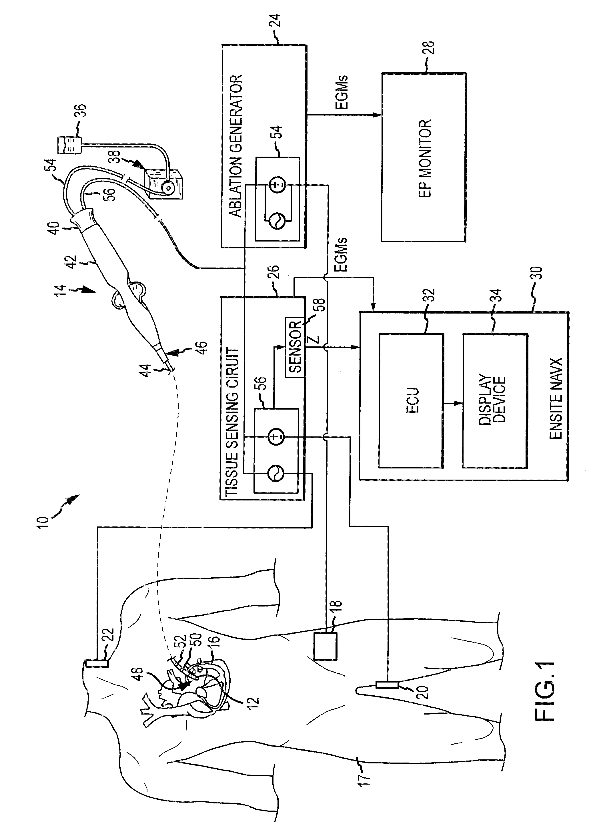
[0029]Referring now to the drawings wherein like reference numerals are used to identify identical components in the various views, FIG. 1 illustrates one embodiment of a system 10 for one or more diagnostic and therapeutic functions including components providing an improved assessment of a degree of coupling between an electrode 12 on a catheter 14 and a tissue 16 in a body 17. In the illustrated embodiment, tissue 16 comprises heart or cardiac tissue. It should be understood, however, that the present invention may be used to evaluate coupling between electrodes and a variety of body tissues. Further, although electrode 12 is illustrated as part of a catheter 14, it should be understood that the present invention may be used to assess a degree of coupling between any type of electrode and tissue including, for example, intracardiac electrodes, needle electrodes, patch electrodes, wet brush electrodes (such as the electrodes disclosed in commonly assigned U.S. patent application S...


PUM


Abstract

A system and method for assessing a degree of coupling between an electrode and tissue in a body is provided. Values for first and second components of a complex impedance (e.g., resistance and reactance or impedance magnitude and phase angle) between the electrode and the tissue are obtained. From these values, a coupling index is calculated that is indicative of a degree of coupling between the electrode and the tissue. The coupling index may be displayed to a clinician in a variety of ways to indicate the degree of coupling to the clinician. The system and method find particular application in ablation of tissue by permitting a clinician to create lesions in the tissue more effectively and safely.

Description

[0001]This application is a continuation-in-part of U.S. patent application Ser. No. 12 / 095,688 filed May 30, 2008, the entire disclosure of which is incorporated herein by reference. U.S. patent application Ser. No. 12 / 095,688 is a national stage application of, and claims priority to, International Application No. PCT / US2006 / 061714 filed Dec. 6, 2006, the entire disclosure of which is incorporated herein by reference. The International Application was published in the English language on Jun. 14, 2007 as International Publication No. WO 2007 / 067941 A2 and itself claims the benefit of U.S. Provisional Patent Application No. 60 / 748,234 filed Dec. 6, 2005, the entire disclosure of which is incorporated herein by reference.BACKGROUND OF THE INVENTION[0002]a. Field of the Invention[0003]This invention relates to a system and method for assessing the degree of coupling between an electrode and tissue in a body. In particular, the instant invention relates to a system and method for asse...

Claims


Application Information

Patent Timeline
Patent Type & Authority Patents(United States)
IPC IPC(8): A61B18/18
CPCA61B5/053A61B18/1492A61B18/1233A61B18/14A61B2018/00178A61B2018/00875A61B2019/465A61B2090/065
Inventor DENO, DON CURTISMILLER, STEPHAN PAULPAUL, SAURAV
Owner ST JUDE MEDICAL ATRIAL FIBRILLATION DIV
Why Eureka
Try Eureka