Method to show a concentration of a contrast agent in a predetermined volume segment by means of tomosynthesis, and corresponding tomosynthesis apparatus

Active Publication Date: 2013-11-26
SIEMENS HEALTHCARE GMBH
View PDF16 Cites 24 Cited by
  • Summary
  • Abstract
  • Description
  • Claims
  • Application Information

AI Technical Summary

Benefits of technology

[0017]The method according to the invention accordingly combines the temporal dual-energy subtraction of tomosynthesis results with the non-temporal dual-energy subtraction of two-dimensional images. As used herein, a dual-energy subtraction is a subtraction of first and second image data, with the first image data being generated with a higher x-ray energy than the second image data. For this reason the method is also called a two-spectra method since the x-ray radiation to generate the low-energy image data and the x-ray radiation to generate the high-energy image data have different wavelengths. Stated differently, the dual-energy subtraction designates the subtraction of a low-energy image from only one high-energy image, both of which are acquired in a short time interval.
[0067]In that the scan speed (i.e. the duration in which the tomosynthesis scan is implemented) is adapted to the contrast agent dynamic, the total radiation dose with which the patient is exposed can be kept optimally low.

Problems solved by technology

In contrast to this, given the generation of the low-energy image the x-ray photons (for the most part) have insufficient energy to strip an electron of a contrast agent atom from the k-shell, such that the x-ray radiation is not absorbed by the contrast agent.

Method used


Image

  • Method to show a concentration of a contrast agent in a predetermined volume segment by means of tomosynthesis, and corresponding tomosynthesis apparatus
  • Method to show a concentration of a contrast agent in a predetermined volume segment by means of tomosynthesis, and corresponding tomosynthesis apparatus
  • Method to show a concentration of a contrast agent in a predetermined volume segment by means of tomosynthesis, and corresponding tomosynthesis apparatus

Examples


Embodiment Construction

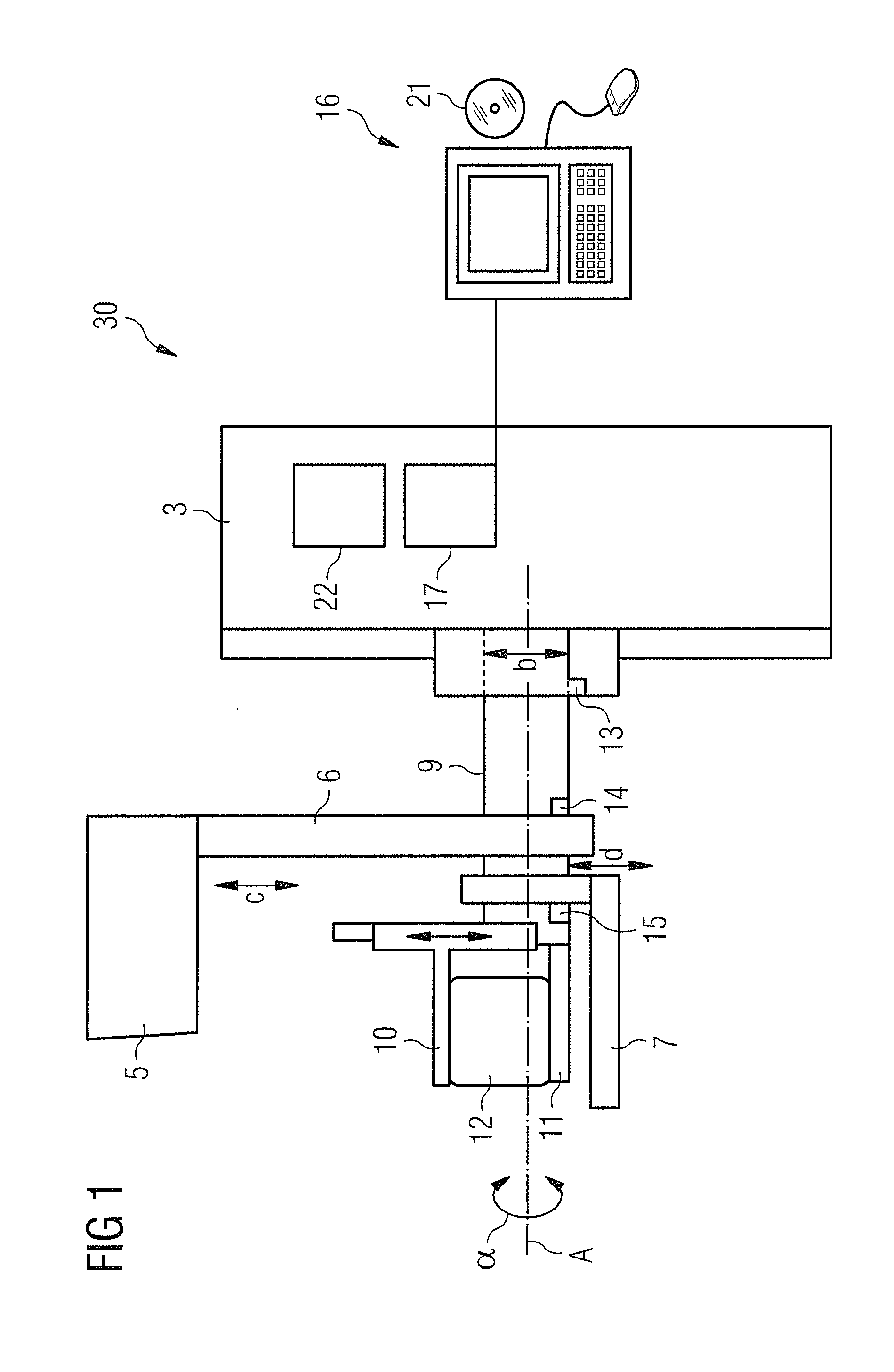
[0082]A tomosynthesis apparatus 30 according to the invention for mammography examinations is schematically shown in FIG. 1. The tomosynthesis apparatus 30 has a support arm 9 that is mounted such that it can pivot in a bearing around a horizontal axis A (see double arrow and angle α). The bearing is arranged on a stand 3 and can be vertically adjusted as indicated with the double arrow b. An arm 6 provided with an x-ray source 5, a flat panel detector 7 and a compression device (consisting of a compression plate 10 and a support plate 11) are arranged on the support arm 9. Schematically shown in FIG. 1 is a female breast 12 compressed by the compression plate 10 and the bearing plate 11. The arm 6 can pivot around the axis A relative to the support arm 1, the detector 7 and the compression device 10, 11. Electromotors 13 through 15 of the tomosynthesis apparatus 30 are provided for height adjustments and pivot motions.

[0083]A control of the tomosynthesis apparatus 30 takes place vi...


PUM


Abstract

In an imaging method and a tomosynthesis apparatus, a two-dimensional low-energy image of the predetermined volume segment is obtained after administration of a contrast agent, followed by a two-dimensional high-energy image and then a high-energy tomosynthesis of the predetermined volume segment is obtained with a high total radiation dose that is significantly higher than the low radiation dose. The two-dimensional low-energy image is subtracted from the two-dimensional high-energy image to generate a result with which the concentration of the contrast agent is visible. Additionally, in a time interval in which an enrichment or a washing-out of the contrast agent occurs within the predetermined volume segment, a tomosynthesis of the predetermined volume segment is automatically implemented to show the concentration of the contrast agent in the predetermined volume segment.

Description

BACKGROUND OF THE INVENTION[0001]1. Field of the Invention[0002]The present invention concerns a method to show a concentration of a contrast agent or the course of the concentration of the contrast agent in a predetermined volume segment by means of a tomosynthesis, as well as a correspondingly designed tomosynthesis apparatus.[0003]2. Description of the Prior Art[0004]To differentiate malignant and benign lesions or tumors, it is known to administer a contrast agent TO the volume segment to be examined and to produce a corresponding evaluation of the lesion or the tumor using the dynamic of the contrast agent concentration. Particularly in the case of malignant tumors, an enrichment of the contrast agent in the tumor (which normally takes place very quickly (within approximately 1 min)) occurs due to the neovascularization. A fast imaging is required in order to measure this increase in the contrast agent concentration. A tomosynthesis scan typically lasts up to 25 s, and when the...

Claims


Application Information

Patent Timeline
Patent Type & Authority Patents(United States)
IPC IPC(8): A61B6/00G06K9/00H05G1/64
CPCA61B6/025A61B6/481A61B6/482A61B6/486A61B6/502
Inventor HOERNIG, MATHIASMERTELMEIER, THOMAS
Owner SIEMENS HEALTHCARE GMBH
Why Eureka
Try Eureka