
Method for diagnosing cancer using lectin

a cancer and lectin technology, applied in the field of cancer diagnosis, can solve the problems of difficult to obtain antibodies for all glycoproteins being discovered on a large scale, many limitations in analysis speed, quantitative reliability, etc., and achieve the effect of easy and quick

Inactive Publication Date: 2014-01-07
KOREA BASIC SCI INST
View PDF3 Cites 0 Cited by
  • Summary
  • Abstract
  • Description
  • Claims
  • Application Information

AI Technical Summary

Benefits of technology

[0036]The present invention provides a method of distinguishing effectively between a normal group and a cancer patient group by analyzing quantitatively marker protein isoforms which have cancer-specific glycan structures which undergo quantitative changes in many kinds of cancer cells. The present invention can diagnose a cancer from a sample of a subject in an easy and quick manner by obtaining information on amounts of marker glycoprotein isoforms having cancer specific glycan structures through a quantitative analysis of marker peptides generated by hydrolysis. And the selected peptides can be useful for a marker for cancer diagnosis.

Problems solved by technology

Therefore, it is necessary to prepare an antibody against antigen glycoprotein and there is a disadvantage that the method cannot be used for proteins of which antibodies cannot be obtained.
In addition, this lectin-blotting method which basically uses a gel-separation technology exhibits many limitations in the analysis speed, quantitative reliability, etc.
However, obtaining reliable antibody is necessary for this sandwich array method and obtaining antibodies for all glycoproteins which are being discovered on a large scale quickly is difficult.
Clinical specimens such as plasma proteins have more than 50,000 components and the concentration of protein components thereof is very dynamic (1˜1011, pg / ml), and therefore, it is very difficult to detect and analyze quantitatively trace amount of plasma biomarker proteins from clinical specimens where high concentration proteins coexist, using liquid chromatography-mass spectrometry (LC / MS / MS) approach (Anderson N. L. et al., Mol.

Method used


Image

  • Method for diagnosing cancer using lectin
  • Method for diagnosing cancer using lectin
  • Method for diagnosing cancer using lectin

Examples


example 1

Sample Preparation

[0102]HCC blood samples (pooled cancer plasma) were prepared by mixing blood samples of ten patients clinically confirmed to have HCC and normal comparative blood samples (pooled control plasma) were prepared by mixing blood samples of ten healthy people whose clinical findings are that there are no cancer-related diseases. Using AAL (aleuria aurantia lectin) which shows a selective affinity for glycoproteins having a fucose glycan, AAL-selective protein samples were isolated from the same amount of blood samples of the HCC patient group and the normal control group. Isolated samples were hydrolyzed, and peptide samples of the HCC patient group and the normal control group were obtained. As for a support for fixing lectins, various kinds of supports, including agarose beads, magnetic beads, etc. may be used. For the analysis of the present clinical blood samples, strepavidine-magnetic beads were used for fixing lectins. That is, respective blood samples of the HCC ...

example 2

Selection of Candidate Markers by Peptide Analysis

[0103]In order to analyze samples prepared in the sample preparation in , LC / ESI-MS / MS was performed using HPLC (high-performance liquid chromatography; trap column: C18, 5 um, 300 um×5 mm; analytical column: C18, 5 um, 75 um×10 cm) tandem LTQ-FT mass spectrometer (Thermo Finnigan), the electrospray ionization (ESI) mass spectrometer. Part of peptide samples prepared by trypsin hydrolysis of each protein sample was diluted 10-fold and 10 μL aliquots were injected into HPLC / mass spectrometer.

[0104]Based on the mass analysis result, hydrolyzed peptides of proteins enriched by AAL can be confirmed through a search engine, such as MASCOT, SEQUEST, etc. Significant proteins were searched from the peptides obtained from LC / ESI-MS / MS analysis and analysis frequency thereof, etc. Glycosylation and cancer-related possibility, etc. of searched proteins were confirmed by investigating protein databases, including Swiss-Prot DB, NCBI nr DB, etc....

example 3

Identification of Marker Peptide using Mass Analysis

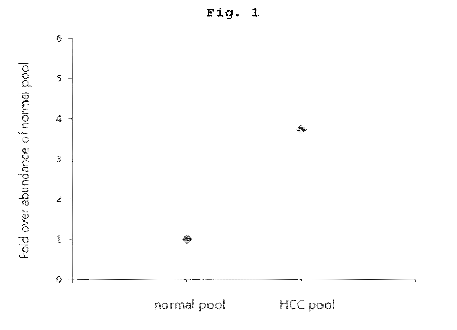
[0108]For MRM quantification of the marker peptide of SEQ ID NO:6 as a representative example among marker peptides of the above [Table 1], isotope-labeled standard of the peptide was prepared and added equally to respective peptide samples of pooled cancer plasma and pooled control plasma which were prepared in as an internal standard for a quantitative analysis. The result of LC / MRM quantitative mass analysis in which the marker peptide of SEQ ID NO:6 was repeatedly quantified for each sample was shown in FIG. 1. That is, it was confirmed that the marker peptide of SEQ ID NO:6 of the HCC patient group was quantitated 3.7 times greater than that of the normal control group.

[0109]When the concentration of target protein is very low in blood, target marker peptide may not be detected directly from the prepared peptide samples according to the above MRM quantitative analysis. In that case, according to a well-known method, quantitat...


PUM

PropertyMeasurementUnit
concentrationaaaaaaaaaa
liquid chromatography-aaaaaaaaaa
massaaaaaaaaaa

Abstract

The present invention relates to a method for diagnosing cancer using information on aberrant glycosylation of glycoproteins, which is related with cancer progression. More particularly, the present invention relates to a peptide marker for cancer diagnosis and a method for diagnosing cancer using the peptide marker, wherein glycoproteins aberrantly glycosylated due to cancer incidence and progression is isolated using lectin; and marker peptides generated by hydrolysis of the glycoproteins isolated by the lectin is selected and quantified.

Description

CROSS-REFERENCES TO RELATED APPLICATION[0001]This patent application claims the benefit of priority from Korean Patent Application No. 10-2012-0031277, filed on Mar. 27, 2012, the contents of which are incorporated herein by reference.REFERENCE TO A SEQUENCE LISTING[0002]This application contains a Sequence Listing submitted electronically as a text file. The text file, named “12fpo-03-09_ST25.txt” has a size in bytes of 3 KB, and was recorded on Apr. 27, 2012. The information contained in the text file is incorporated herein by reference in its entirety pursuant to 37 CFR §1.52(e)(5).BACKGROUND OF THE INVENTION[0003]1. Field of the Invention[0004]The present disclosure relates to a method for diagnosing cancer by isolating and enriching glycoproteins which are aberrantly glycosylated due to cancer by using lectin, selecting polypeptides, and quantitatively analyzing the polypeptides.[0005]2. Description of the Related Art[0006]Protein glycosylation is one of the most representative...

Claims


Application Information

Patent Timeline
Patent Type & Authority Patents(United States)
IPC IPC(8): G01N33/53
CPCG01N33/6818G01N2400/02G01N2333/42G01N33/6842G01N33/57484G01N33/57438G01N2030/027
Inventor AHN, YEONG HEEYOO, JONG SHINSHIN, PARK MINJI, EUN SUN
Owner KOREA BASIC SCI INST
Learn More