VLPs derived from cells that do not express a viral matrix or core protein

a cell and core protein technology, applied in the field of vlps, can solve the problems of viral replication and the inability of recombinant subunit protein vaccines to elicit protective immunity

Active Publication Date: 2014-04-15
NOVAVAX
View PDF6 Cites 6 Cited by
  • Summary
  • Abstract
  • Description
  • Claims
  • Application Information

AI Technical Summary

Problems solved by technology

However, results from Phase II studies conducted at several clinical sites in human volunteers vaccinated with several doses of influenza vaccines comprised of HA and / or NA proteins indicated that the recombinant subunit protein vaccines did not elicit protective immunity.
However, in spite of the fact that both full-length M2 protein and M2-HBcAg particles induced detectable antibodies and protection in mice, it was unlikely that future influenza vaccines would be based exclusively on the M2 protein, as the M2 protein was present at low copy number per virion, was weakly antigenic, was unable to elicit antibodies that bound free influenza virions, and was unable to block virus attachment to cell receptors (i.e. virus neutralization).
VLPs are structurally similar to mature virions, but lack the viral genome making it impossible for viral replication to occur.

Method used


Image

  • VLPs derived from cells that do not express a viral matrix or core protein
  • VLPs derived from cells that do not express a viral matrix or core protein
  • VLPs derived from cells that do not express a viral matrix or core protein

Examples


example 1

VLP Formation Induced by Expression of Influenza HA and NA Proteins

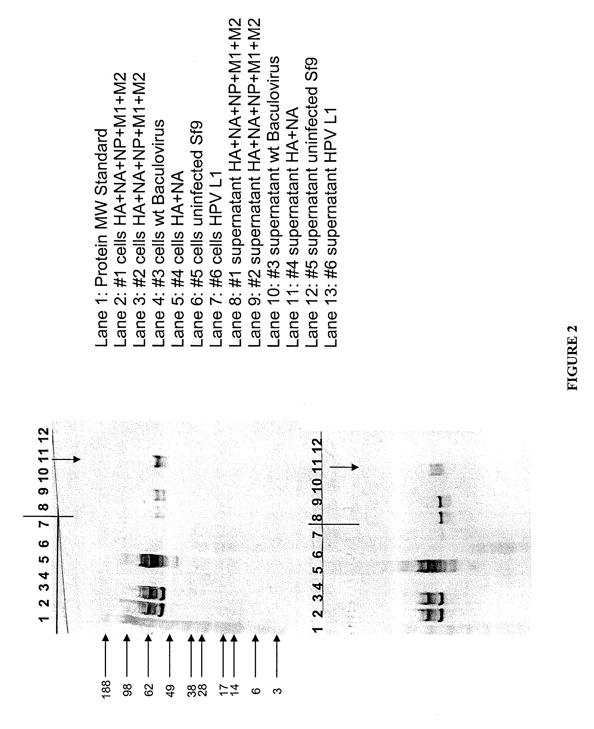
[0119]Influenza A / Sydney / 5 / 97 (H3N2) virus HA, NA M1, M2, and NP genes were expressed in Spodoptera frugiperda cells (Sf-9S cell line; ATCC PTA-4047) using the baculovirus bacmid expression system. Infection of permissive Sf-9S insect cells with recombinant baculovirus resulted in co-expression of all five influenza genes or subcombinations thereof in each Sf-9S cell infected with such recombinant baculovirus. The samples, as defined below, are the influenza proteins expressed in a single cell or controls. HPV 16L1, a protein from human papilloma virus, is used as a control.

Samples:

1. HA+NA+NP+M1+M2

2. HA+NA+NP+M1+M2

3. wt baculovirus

4. HA+NA

5. uninfected Sf9 cells

6. HPV 16L1

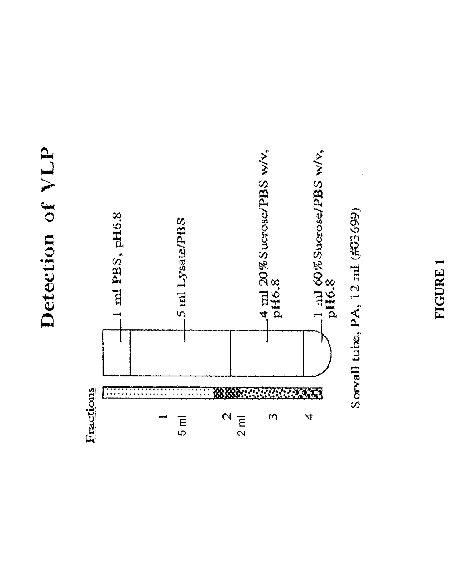
[0120]After growing the infected cells for four days, the cells were pelleted and frozen at −80° C. The cells were thawed at room temperature, resuspended in 4 ml of PBS, pH6.8 and transferred into 560 ml tubes. The cells were sonicated and clarifi...

example 2

Expression of Single Influenza Membrane Proteins Induces VLP Formation

[0126]Sf9 cells were infected with recombinant baculovirus expressing various influenza proteins or combinations thereof. Infected cells were incubated in serum-free medium. The media supernatants were pelleted through 20% sucrose cushion. Pellets were resuspended and subjected to western blot analysis. The blot was probed using a polyclonal antibody specific for influenza virus (FIG. 7).

Lanes:

1. Molecular weight standard

2. HA+NA+NP+M1+M2

3. HA+NA+M1+M2

4. HA

5. NA

6. M1

7. M2

[0127]Immunoreactive bands are observed in all lanes, but lane 7. These data show that influenza proteins are present in the material, likely VLPs, which could be purified by pelleting through sucrose cushion. Therefore, the supernatant from cells infected with baculovirus expressing either HA protein only, NA protein only or M1 protein only exhibited evidence of VLP formation.

example 3

Expression of Chimeric Proteins Derived from Influenza Membrane Proteins Drives VLP Formation

[0128]Sf9 cells were infected with recombinant baculovirus expressing chimeric proteins. The chimeric proteins contained enhanced green fluorescent protein (eGFP) fused to either the cytoplasmic tail of influenza M2 protein or the cytoplasmic tail of influenza HA protein as illustrated in FIG. 8. Supernatants of the infected cells were purified through a sucrose gradient and subjected to SDS-PAGE and western blot analyses. The resulting coomassie-stained gel is shown in FIG. 9A and the corresponding western blot is shown in FIG. 19B. The blot in FIG. 9B was probed with a polyclonal antibody against GFP. The immunoreactive bands present in lanes 2 and 4 indicate the presence of chimeric proteins (eGFP-M2 cytoplasmic tail and eGFP-HA cytoplasmic tail, respectively) in the supernatant samples purified by sucrose gradient. These data suggest that VLPs are formed in cells expressing the chimeric ...


PUM

PropertyMeasurementUnit
pHaaaaaaaaaa
pHaaaaaaaaaa
temperatureaaaaaaaaaa

Abstract

The present invention discloses novel influenza virus-like particles (VLPs) that contain chimeric proteins or influenza membrane proteins. The chimeric proteins are derived from fragments of influenza membrane proteins fused to heterologous proteins. The invention includes antigenic formulations and vaccines comprising VLPs of the invention as well as methods of making and administering VLPs to vertebrates, including methods of inducing immunity to infections, such as influenza.

Description

CROSS-REFERENCE TO RELATED APPLICATIONS[0001]This application claims priority to U.S. provisional application 60 / 940,201, filed May 25, 2007, which is herein incorporated by reference in its entirety.DESCRIPTION OF THE TEXT FILE SUBMITTED ELECTRONICALLY[0002]The contents of the text file submitted electronically herewith are incorporated herein by reference in their entirety: A computer readable format copy of the Sequence Listing of the Sequence Listing (filename: NOVV 017 01US SeqList_ST25.txt, date recorded: May 27, 2008, file size 7 kilobytes).BACKGROUND[0003]Influenza virus is a member of the Orthomyxoviridae family, and can be further classified into three subtypes: A, B, and C (for review, see Murphy and Webster, (1996) Virology, Vol. 1, pp. 1397-1444. Lippincott-Raven, Philadelphia). Influenza subtype A causes the most severe disease in humans. The A strain can be subdivided into different serotypes according to which forms of two surface antigens (hemagglutinin and neuramin...

Claims


Application Information

Patent Timeline
Patent Type & Authority Patents(United States)
IPC IPC(8): C12N15/44A61K39/145
CPCA61K2039/5258C12N2760/16123C12N2710/16723C07K2319/00A61K39/145C12N7/00A61P31/16
Inventor SMITH, GALEPUSHKO, PETERMAHMOOD, KUTUBZHAO, BIN
Owner NOVAVAX
Why Eureka
Try Eureka