Medical image display processing method, device, and program
a medical image and display processing technology, applied in image enhancement, instruments, applications, etc., can solve the problem of placing a large and achieve the effect of reducing the burden on the physician during interpretation and easy and reliably extraction
- Summary
- Abstract
- Description
- Claims
- Application Information
AI Technical Summary
Problems solved by technology
Method used
Image
Examples
first embodiment
[0053]Next, basic processing procedures applied to an image display processing method of a first embodiment according to the present invention that is performed by the medical image display processing device will be described with reference to a flowchart shown in FIG. 3. In this flowchart, each ellipse represents an image or a processed product.
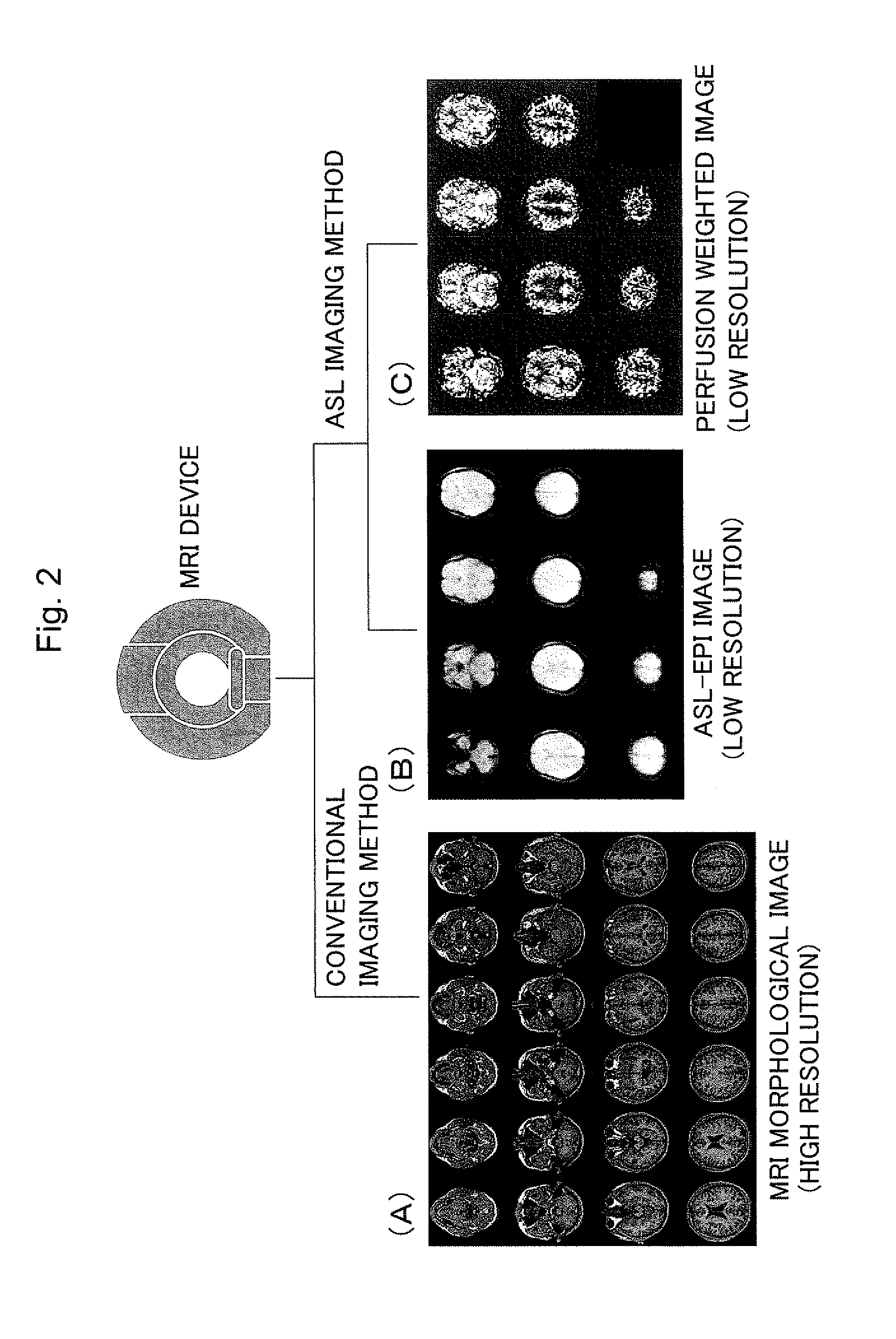
[0054]In the present embodiment, perfusion weighted images and ASL-EPI images are inputted as input images.
[0055]First, the two types of inputted images are subjected to image reconstruction (step 1). The image reconstruction is a process, in which the two types of images are re-sampled three-dimensionally and divided into voxels with equal size in all directions to make the units of the three-dimensional voxels, corresponding to pixels in two-dimensions, of the two types of images the same size.
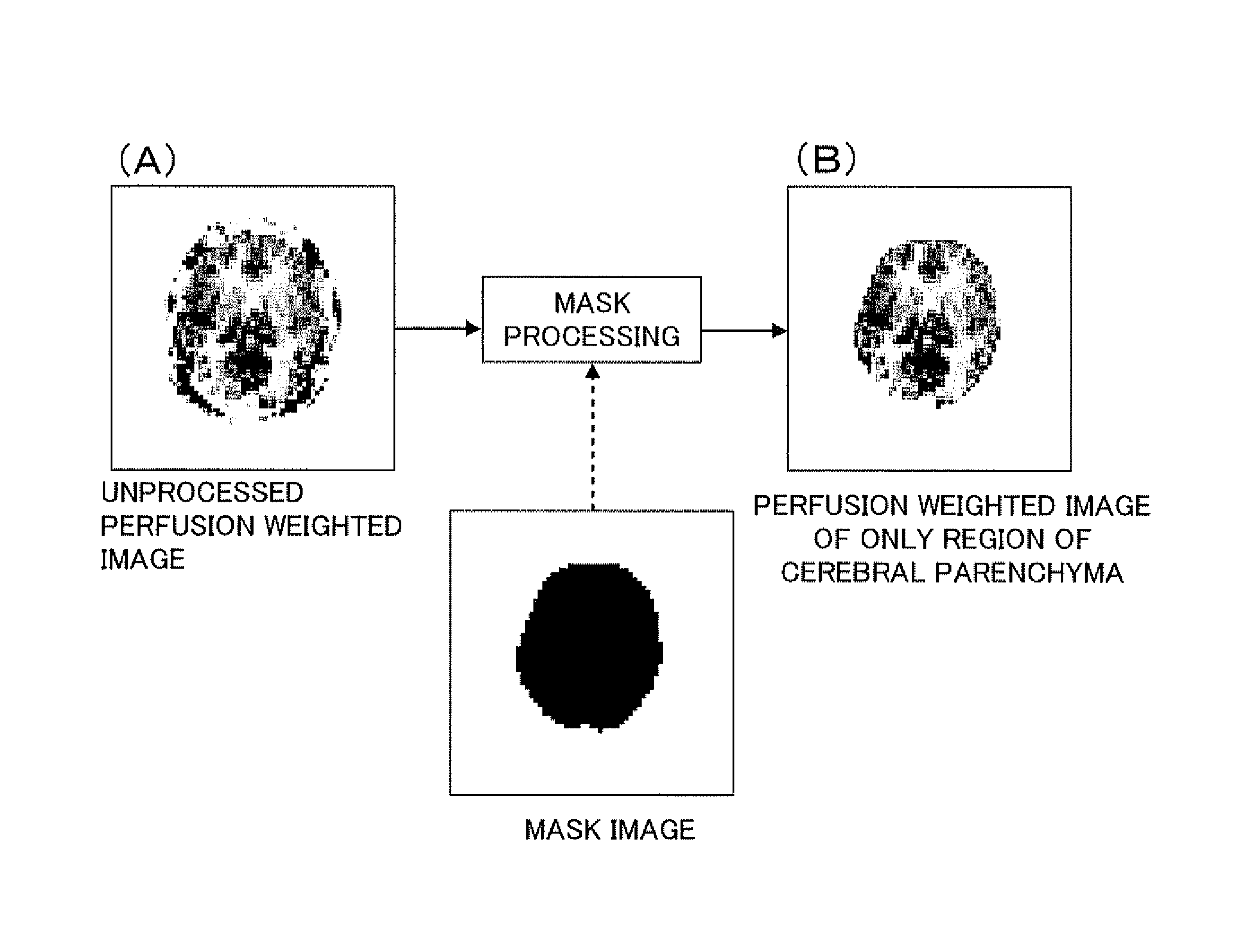
[0056]Next, mask images are produced from the ASL-EPI images by mask production (step 2).
[0057]FIG. 4 shows a mask image (B) produced by binarizing ...
second embodiment
[0076]Next, an image display processing method of a second embodiment will be described with reference to a flowchart shown in FIG. 10.
[0077]In the present embodiment, as in the basic processing procedures, perfusion weighted images and ASL-EPI images are used as input images, and the same processing as in the above steps 1 to 4 is used to produce perfusion weighted images of only the region of the cerebral parenchyma (steps 21 to 23). However, the processing related to the determination of mask boundaries is omitted from the figure.
[0078]In the present embodiment, each of the perfusion weighted images in the subject brain coordinate system A that have been produced in step 23 is transformed into an image in the standard brain coordinate system. First, a spatial transformation field used to transform an ASL-EPI image into an image on the standard brain is estimated using spatial normalization in which the ASL-EPI image is fitted to a T1 weighted image serving as a template and store...
third embodiment
[0088]Next, an image display processing method of a third embodiment will be described with reference to a flowchart shown in FIG. 13.
[0089]In the present embodiment, perfusion weighted images and ASL-EPI images are used as input images, and the process until image reconstruction in step 31 is the same as that in the second embodiment. However, transformation fields to the standard brain are first formed from the reconstructed ASL-EPI images using spatial normalization in the same manner as in the above step 24 (step 32), and the transformation fields are applied to the perfusion weighted images before mask processing to transform them into images in the standard brain coordinate system (step 33).
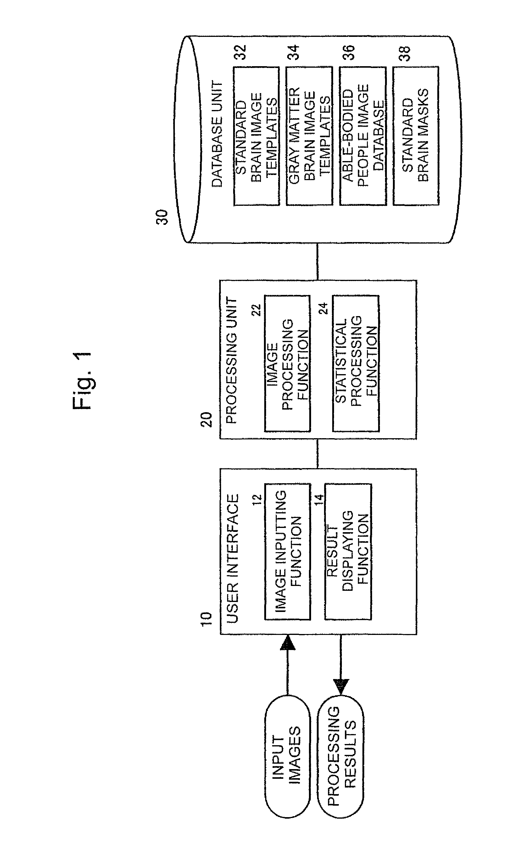
[0090]Next, the perfusion weighted images transformed into the standard brain are subjected to mask processing using the known standard brain masks 38 stored in the database unit 30 (step 34).
[0091]Although not illustrated, perfusion weighted images of only the region of the cerebral parenc...
PUM
 Abstract
Description
Claims
Application Information
 - R&D
- Intellectual Property
- Life Sciences
- Materials
- Tech Scout
- Unparalleled Data Quality
- Higher Quality Content
- 60% Fewer Hallucinations
Browse by: Latest US Patents, China's latest patents, Technical Efficacy Thesaurus, Application Domain, Technology Topic, Popular Technical Reports.
© 2025 PatSnap. All rights reserved.Legal|Privacy policy|Modern Slavery Act Transparency Statement|Sitemap|About US| Contact US: help@patsnap.com


