Supercharge Your Innovation With Domain-Expert AI Agents!

Monoclonal antibody against necrosis marker PRDX4 and use thereof

a monoclonal antibody and necrosis technology, applied in the field of new necrosis markers, can solve the problems of limited prior art techniques for detecting necrosis focus biomarkers

Active Publication Date: 2015-10-20
FUJIFILM CORP
View PDF24 Cites 0 Cited by
  • Summary
  • Abstract
  • Description
  • Claims
  • Application Information

AI Technical Summary

Benefits of technology

The patent is about a new marker for necrosis, which can be used to detect areas of necrosis in various diseases, such as cancer, heart disease, and inflammation. The marker is a protein that can be specifically stained with an antibody, making it a useful tool for detecting foci of necrosis in human tissues. The antibody can also be labeled with an isotope to allow for specific imaging of necrotic areas in mouse models. Overall, this patent provides a valuable tool for studying and diagnosing various diseases and potentially improving treatment outcomes.

Problems solved by technology

However, prior art techniques concerning biomarkers for detecting a focus of necrosis are limited to the three kinds of antibodies selected by using insoluble intracellular antigens derived from a lymphoma cell line or lung cancer cell line (Patent document 1), an antibody against a soluble nuclear matrix protein (Patent document 2), an antibody or low molecular weight antibody specifically binding to the nucleus extract identified as a center of necrosis of a tumor and histone H1 (Patent document 3), and so forth.

Method used


Image

  • Monoclonal antibody against necrosis marker PRDX4 and use thereof
  • Monoclonal antibody against necrosis marker PRDX4 and use thereof
  • Monoclonal antibody against necrosis marker PRDX4 and use thereof

Examples


example 1

Preparation of Necrotic Focus Cell Model System

[0076]The method of the present invention requires a cell in which necrosis is induced as a cell model forming a focus of necrosis. Therefore, necrosis was induced in the cells of cervical cancer cell strain, HeLa cells, to prepare a cell model forming a focus of necrosis. The HeLa cells are generally adhesive cells, and 2×10⁵ to 1×10⁶ cells are cultured on a 10-cm2 petri dish in a scaffold-dependent manner. In usual culture, they are cultured at 37° C. and a cell density of 2×10⁴ to 1×10⁵ cells / ml in 10 ml of blood serum-containing DMEM medium (Dulbecco's modified Eagle medium) under a gas phase of 5% CO2 and 95% air and saturated humidity. The HeLa cells have high proliferation potency, and in the highly nutritious blood serum-containing DMEM medium, they proliferate to reach confluent in a 10-cm2 petri dish within two to three days. Since the HeLa cells are cells of a cancer cell line, even if the cells reach confluent in a petri dis...

example 2

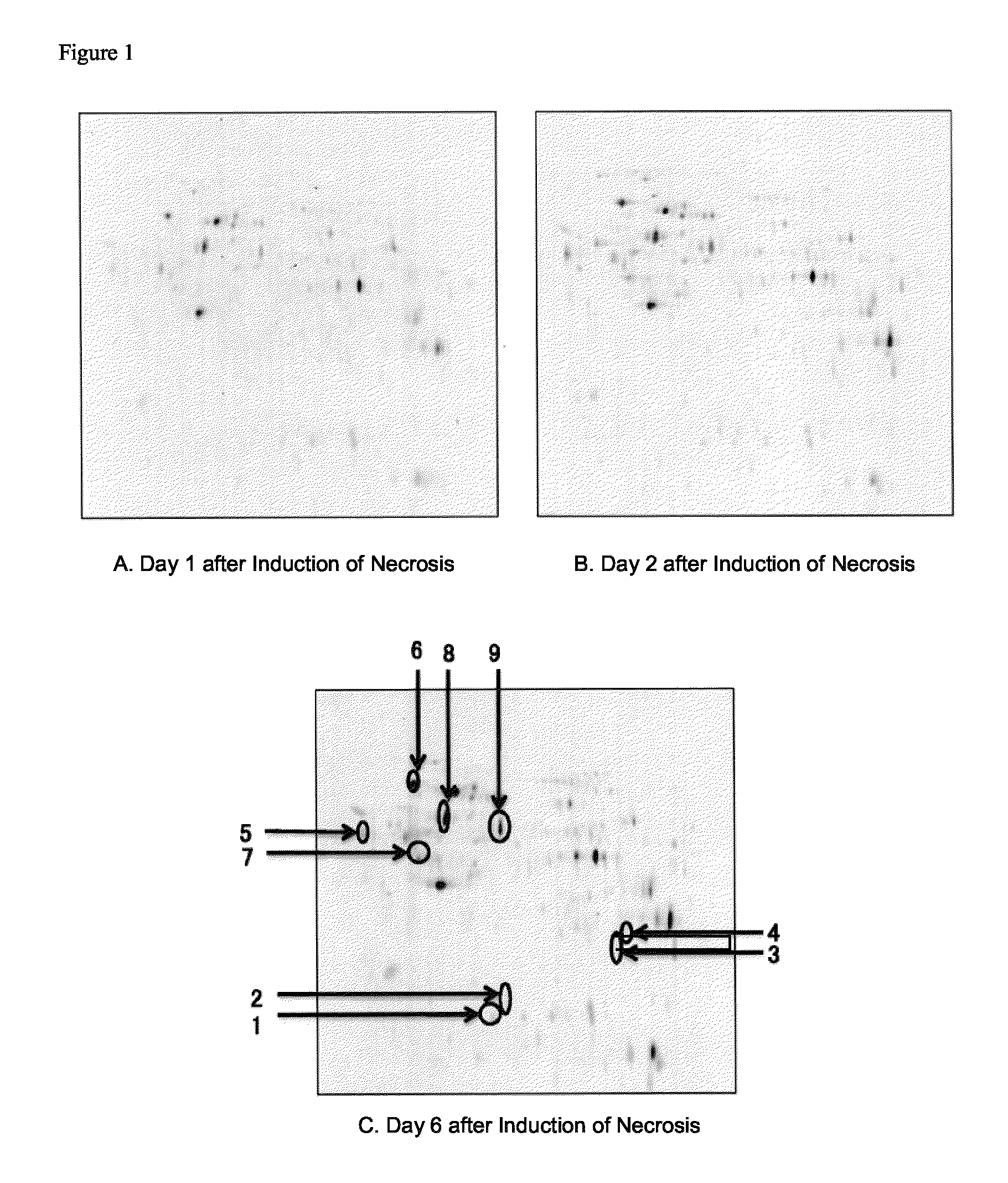
Identification of Marker Molecules Specific to Focus of Necrosis

[0085]2.1 Preparation of Cell Extracts

[0086]The HeLa cells on the day 1 to day 6 after the induction of necrosis in the DMEM medium containing 10% FCS and the HeLa cells cultured in the usual manner obtained in Example 1 were collected, and washed twice with ice-cooling PBS(−). An ice-cooled solubilization buffer (20 mM Tris-HCl (pH 7.5), 150 mM NaCl, 1 mM EDTA, 1% NP-40) in a volume of 500 to 1,000 μl was added to the cells to suspend the cells, and the suspension was transferred to a 1.5-ml tube. The soluble fraction of the cells was extracted with rotating the tube at 4° C. for 30 minutes, and centrifuged at 15,000 rpm for 15 minutes, and the supernatant was collected as a cell extract. The collected cell extract was purified by using 2D-Clean up kit (80-6484-51, Amersham) according to the attached protocol, and dissolved in an appropriate volume of a lysis buffer (30 mM Tris (pH 8.5) 7 M urea, 2 M thiourea, 4% CHAPS...

example 3

Preparation of Monoclonal Antibodies

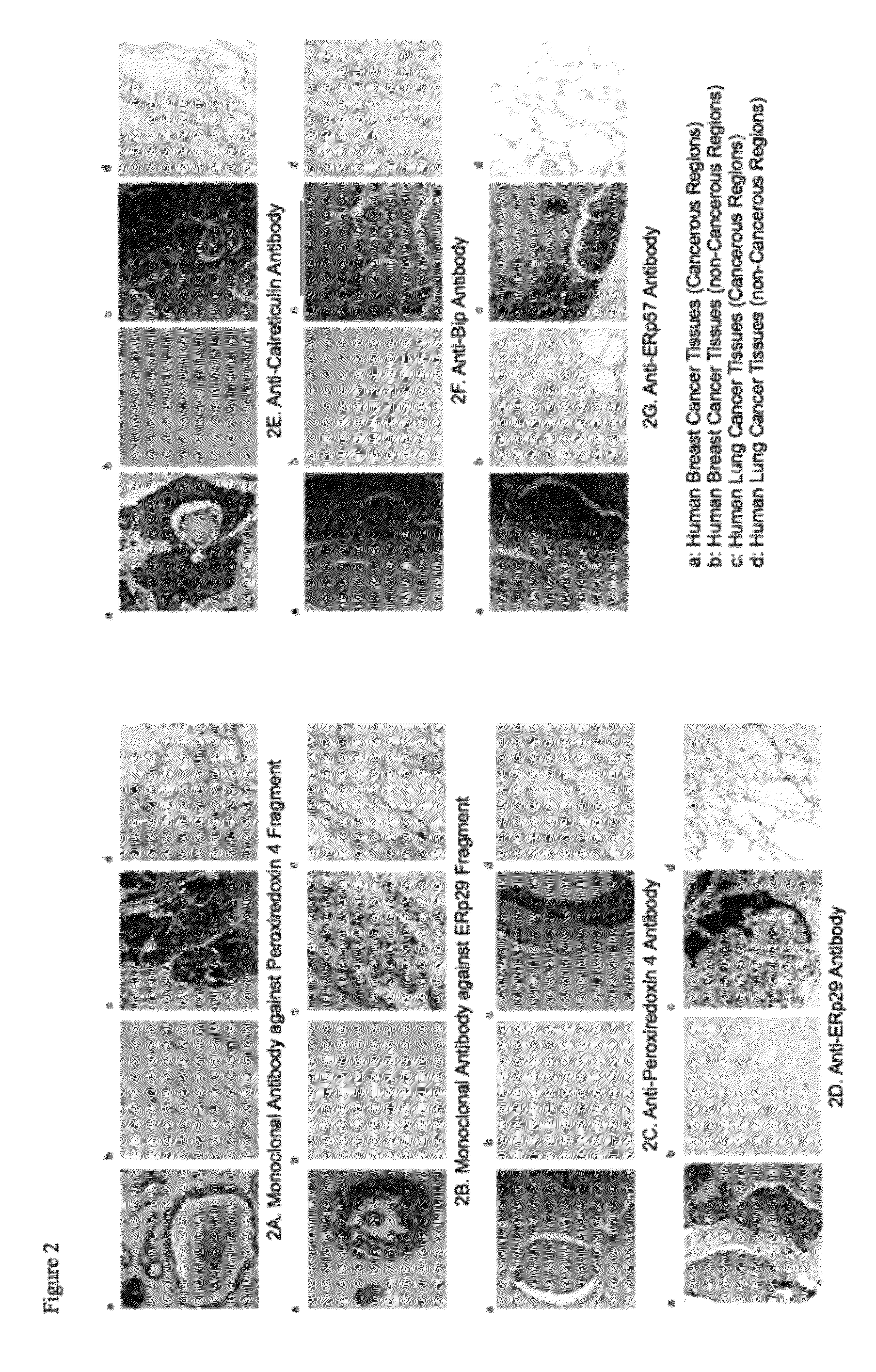
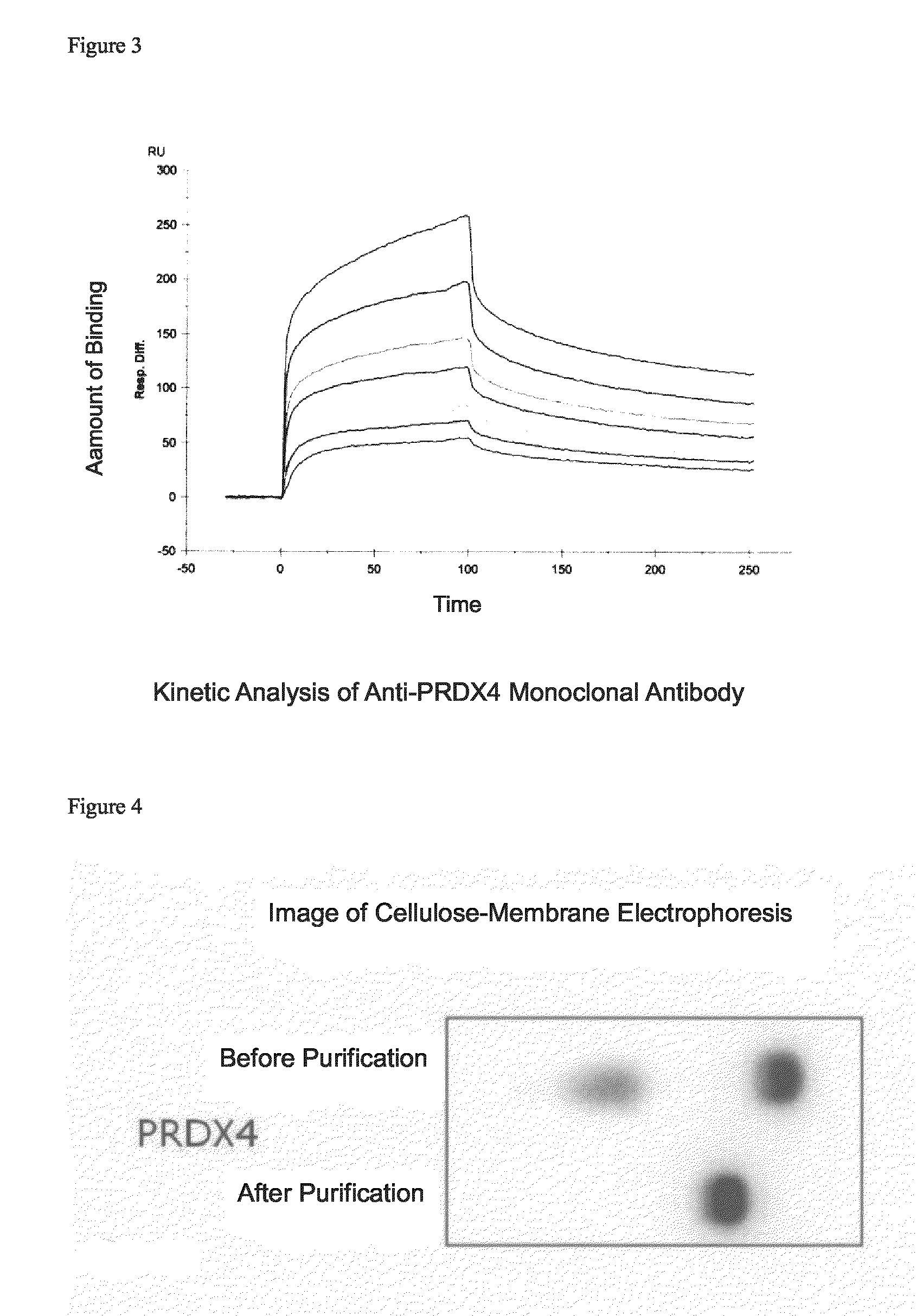
[0109]3.1 Sensitization of Immunized Animal and Cell Fusion

[0110]For the peroxiredoxin 4 fragment and the ERp29 fragment among the proteins identified as necrotic focus-specific markers, mouse monoclonal antibodies that could recognize each sequence were prepared on the basis of the N-terminal amino acid sequences identified in Example 2.

[0111]A peptide of 10 residues consisting of the aforementioned N-terminus 9 amino acid residues of the peroxiredoxin 4 fragment or the ERp29 fragment and a cysteine residue added to the C-terminus of the foregoing residues for binding with a carrier polymer (KLH) was chemically synthesized in a conventional manner in an amount of about 10 mg, and purified by HPLC, and the purified peptide was used as an antigen peptide. The antigen peptide was crosslinked with KLH in a conventional manner, and mixed with an equal amount of the Freund's complete adjuvant (RM606-1, Mitsubishi Chemical Iatron), and the mixture was e...


PUM


Abstract

[PROBLEM] To provide a monoclonal antibody against a biomarker which shows high specificity and can be effectively used in detection and diagnosis of various lesions relevant to various kinds of carcinomas and foci of necrosis, and so forth.[MEANS] A monoclonal antibody against a necrosis marker consisting the following amino acid sequence: (1) the amino acid sequence of SEQ ID NO: 1, or (2) an amino acid sequence having substitution, deletion and / or insertion of one or several amino acid residues in the amino acid sequence of (1) or sharing a homology of 90% or more with the amino acid sequence of (1), and showing the same function, activity or property as that of the amino acid sequence of (1) as a protein.

Description

CROSS REFERENCE TO RELATED APPLICATIONS[0001]This application is a National Stage of International Application No. PCT / JP2011 / 055356 filed Mar. 8, 2011, claiming priority based on Japanese Patent Application No. 2010-050106 filed Mar. 8, 2010, the contents of all of which are incorporated herein by reference in their entirety.TECHNICAL FIELD[0002]The present invention relates to a novel necrosis marker, a method for detecting a focus of necrosis using amount of the necrosis marker, a kit for detecting a focus of necrosis containing a compound that specifically reacts with the necrosis marker, a composition for diagnostic imaging or conjugate for a therapeutic treatment comprising a compound that specifically reacts with the necrosis marker and a labeling compound or a compound effective for a therapeutic treatment, a kit for diagnostic imaging or pharmaceutical composition containing the conjugate, a method for identifying a necrosis marker, which comprises detecting necrosis releva...

Claims


Application Information

Patent Timeline
Patent Type & Authority Patents(United States)
IPC IPC(8): G01N33/53G01N33/574A61K51/10C07K16/30C07K16/18C07K16/00
CPCG01N33/574A61K51/1045C07K16/18C07K16/30G01N2800/00A61P1/16A61P1/18A61P9/00A61P9/10A61P21/00A61P35/00A61K51/1093C07K2317/14C07K2317/76C07K2317/92G01N33/57496G01N2333/47G01N2333/908
Inventor SUGANO, SUMIOKANZAKI, YUKARISAGA, TSUNEOTSUJI, ATSUSHI
Owner FUJIFILM CORP
Features
  • R&D
  • Intellectual Property
  • Life Sciences
  • Materials
  • Tech Scout
  • Unparalleled Data Quality
  • Higher Quality Content
  • 60% Fewer Hallucinations
Learn More