
Surgical clips for laparoscopic procedures

a technology for surgical clips and laparoscopic procedures, applied in the field of surgical ligation clips, can solve problems such as patient blood loss, interference on the surgical site, and damage to nearby tissu

Active Publication Date: 2017-03-21
CONMED CORP
View PDF126 Cites 67 Cited by
  • Summary
  • Abstract
  • Description
  • Claims
  • Application Information

AI Technical Summary

Problems solved by technology

Movement or slippage of the surgical clip or failure to fully close a blood vessel may cause one or more of the following: damage to nearby tissue, interference on the surgical site, patient blood loss, a lethal drop in blood pressure, or loss of the clip inside the patient.
Surgical clips that cause tissue or blood vessel damage may result in internal bleeding, a lethal drop in blood pressure, infections, or longer recovery periods.
These previously disclosed surgical clips do not adequately securely fasten to and completely close a blood vessel while minimizing surrounding tissue damage.
Another problem for these previously disclosed surgical clips is the phenomenon known as the “scissoring” effect
This misalignment causes the surgical clip to cut or sever blood vessels during application.

Method used


Image

  • Surgical clips for laparoscopic procedures
  • Surgical clips for laparoscopic procedures
  • Surgical clips for laparoscopic procedures

Examples


Embodiment Construction

[0060]While several variations of the present invention have been illustrated by way of example in particular embodiments, it is apparent that further embodiments could be developed within the spirit and scope of the present invention, or the inventive concept thereof. However, it is to be expressly understood that such modifications and adaptations are within the spirit and scope of the present invention, and are inclusive, but not limited to the following appended claims as set forth.

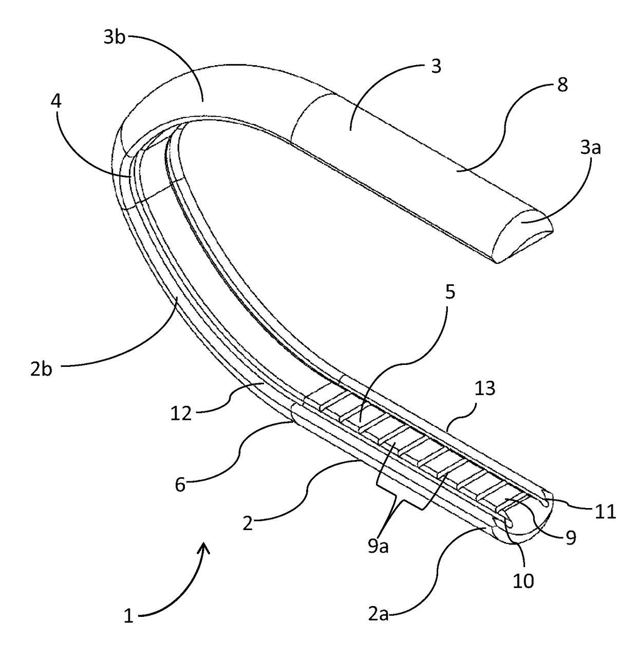
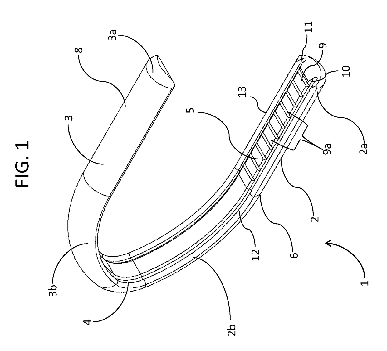
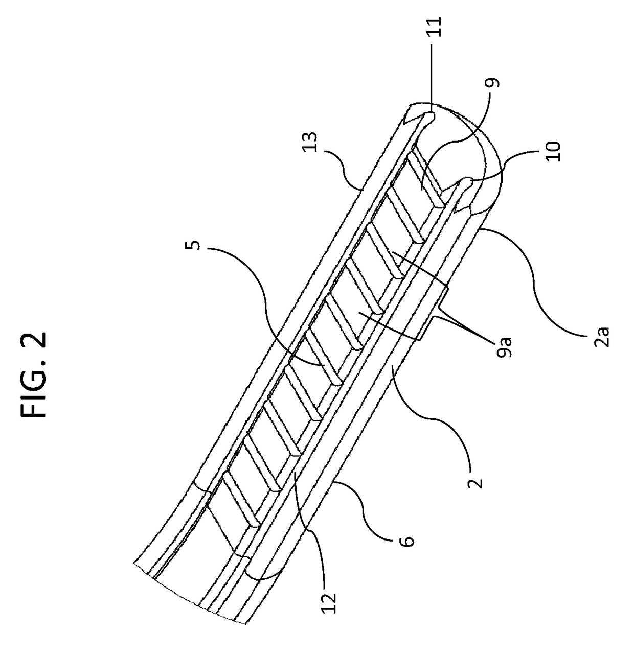
[0061]The subject invention comprises a surgical vessel ligating clip 1, as shown in FIGS. 1-10 and 12-14.

[0062]FIGS. 1, 3, 5 and 6 illustrate the surgical vessel ligating clip 1 in the open or uncompressed position. The surgical vessel ligating clip 1 will be in an open position before attachment or application over a blood vessel to ligate or occlude that vessel. FIGS. 8-10 illustrate the closed or compressed position of surgical vessel ligating clip 1 alone and clamped over a tubular vessel V.

[0063...


PUM


Abstract

A surgical clip for a clip applier to ligate vessels. The clip contains two opposing legs connected in parallel by an apex to form a U-shape. Each leg has an outer surface with a half-round cross-sectional configuration for minimizing tissue damage by preventing clip scissoring during ligation. Each leg has an inner surface with a flat central surface, two concave grooves on opposing sides of the central surface, and two convex ridges on opposing sides of the each groove. As a vessel is ligated, vessel tissue moves into the concave grooves while remaining frictionally engaged by the compressive forces applied by the central surfaces and convex ridges of each leg so that the clip does not move on the vessel. The edges of the grooves and the edges of the ridges of each leg are rounded to prevent any cutting of the ligated vessel.

Description

CROSS REFERENCE TO RELATED APPLICATION[0001]This application claims the benefit of U.S. Provisional Patent Application No. 61 / 312,505 filed on Mar. 10, 2010, the contents of which are incorporated herein by reference.FIELD OF THE INVENTION[0002]This invention relates to novel surgical ligation clips for ligating blood vessels during laparoscopic or endoscopic procedures.BACKGROUND OF INVENTION[0003]In order to operate on a given tissue or a blood vessel, surgeons must ligate or occlude nearby blood vessels to prevent patient blood loss.[0004]Since laparoscopic and endoscopic surgical procedures are conducted through a small incision in the skin or natural body orifices, the surgeon must have long and narrow tools to complete this ligation or occlusion of blood vessels. These tools must be small enough to be inserted through an incision and long enough to reach the desired blood vessels within the patient.[0005]Surgeons employ small surgical clips and long clip appliers to ligate or ...

Claims


Application Information

Patent Timeline
Patent Type & Authority Patents(United States)
IPC IPC(8): A61B17/122A61B17/128
CPCA61B17/122A61B17/1285A61B17/12A61B17/08A61B2017/00004A61B2017/00778
Inventor MENN, PAVEL
Owner CONMED CORP
Learn More