

Quantitative in situ characterization of heterogeneity in biological samples
a heterogeneity and quantitative in situ technology, applied in the field of cell profiling of biological samples, can solve the problems of limited sample amount, loss of original location information, and limited ability to determine relative characteristics of targets
- Summary
- Abstract
- Description
- Claims
- Application Information
AI Technical Summary
Problems solved by technology
Method used
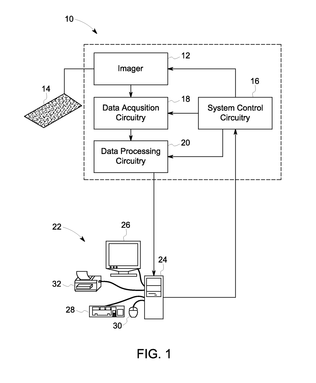
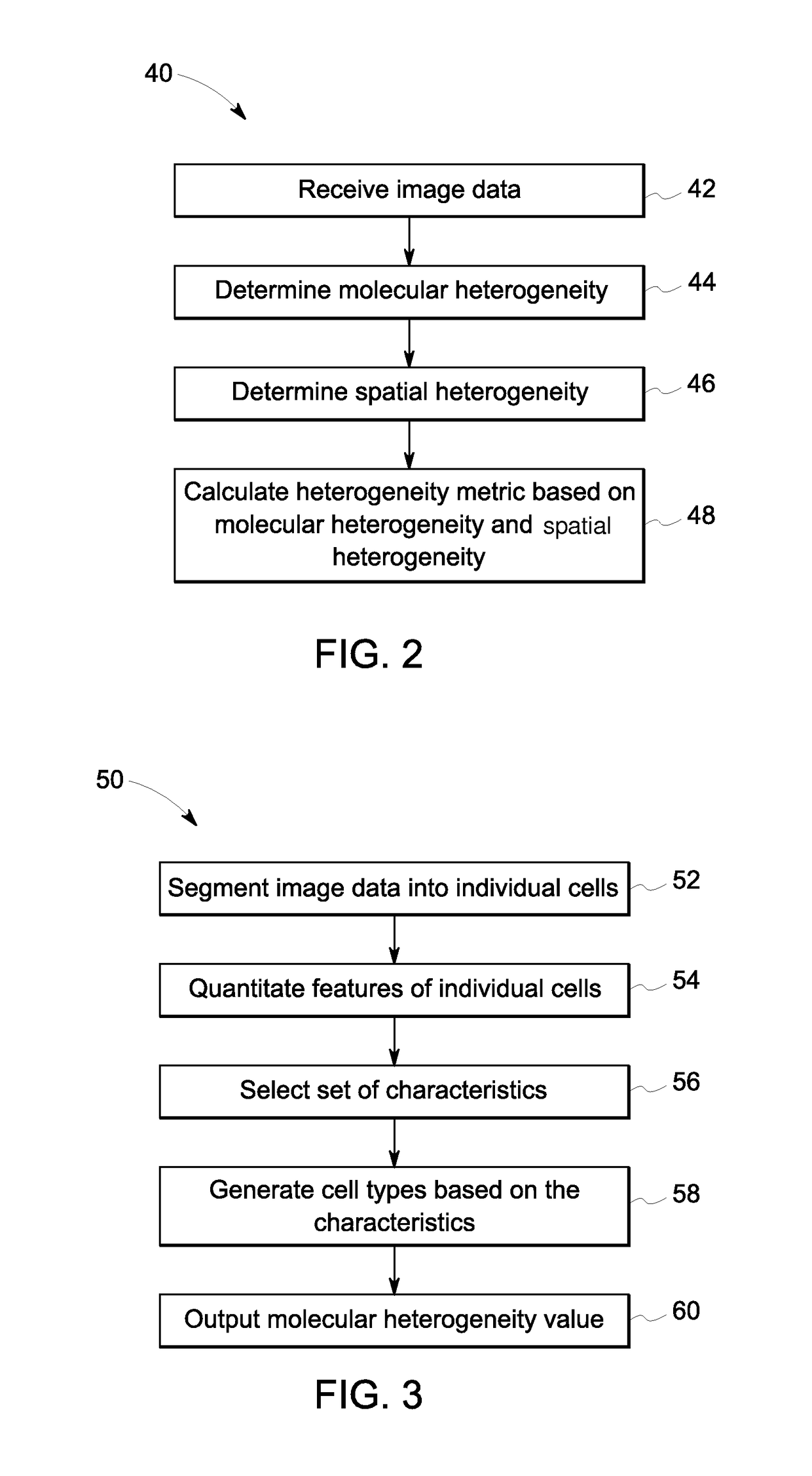
Image

Examples
examples
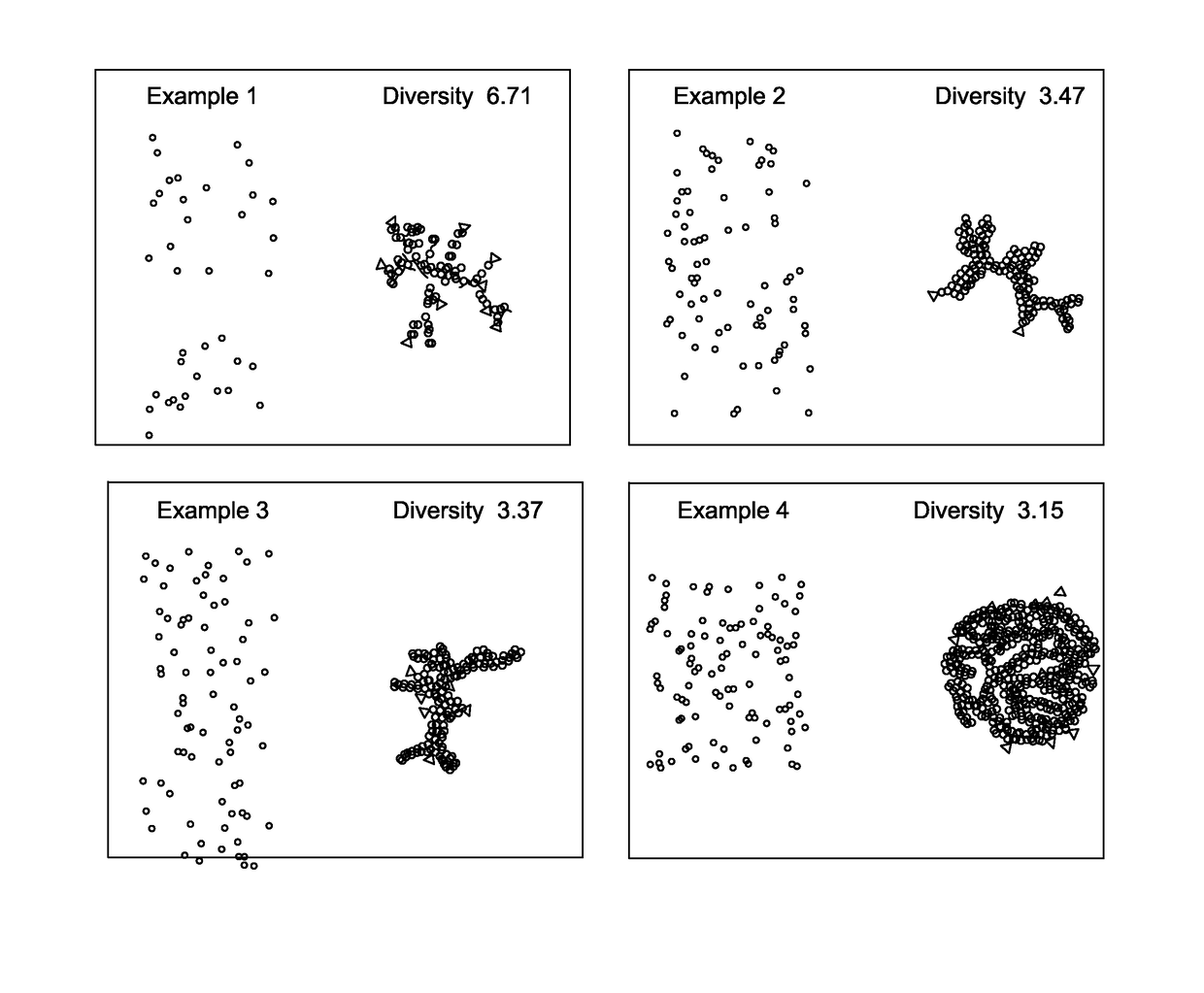
[0054]Tissue samples of colorectal cancer were collected at the Clearview Cancer Institute of Huntsville Ala. and provided to GE Global Research by Clarient Inc. This tissue microarray (TMA) imaging cohort consisted of 747 paraffin-embedded patient tumor samples distributed across three slides. These samples underwent multiplexed fluorescence microscopy (MxIF) and the results and experimental details have been reported previously (Gerdes 2013). Clinical measures for each patient were provided including the histological tumor grade, cancer stage, patient sex, age, chemo treatment (yes / no), and follow-up monitoring of 10 years (medium of 4.1 years). A total of 692 samples passed MxIF image quality assessments. Table 1 presents a breakdown of samples by histological grade and cancer stage. Table 2 presents the number of patients with or without a reoccurrence event during follow-up, broken down by cancer stage and treatment protocol. For each tissue sample (i.e. field of view (FOV)), t...
PUM
 Abstract
Description
Claims
Application Information