Tubular bioengineered smooth muscle structures

a technology of smooth muscle and tubular structure, which is applied in the direction of skeletal/connective tissue cells, ligaments, prostheses, etc., can solve the problems of inability to generate any propulsive force, risk of blockage, and inability to adapt to artery replacemen

Active Publication Date: 2017-06-13
WAKE FOREST UNIV HEALTH SCI INC
View PDF6 Cites 8 Cited by
  • Summary
  • Abstract
  • Description
  • Claims
  • Application Information

AI Technical Summary

Problems solved by technology

They often fail to generate any propulsive force, and function only as conduits -- with some risk of blockage.
Such artificial blood vessels may be suitable for vein replacement but have not been shown to be suitable to artery replacement.
Any slowdown in this propulsive, peristaltic movement, such as localized or segmental paralysis (referred to as “paresis”), can result in localized or segmental inertia that is detrimental for the proper nutrition and can lead to obstruction, infection or morbidity.
Surgical interventions to remedy long-gap esophageal atresia are often marred by dysmotility and impaired quality of life.
This becomes challenging due to the phenomenon of at-will “primary peristalsis,” a complex interplay between the central and enteric nervous systems.
Surface functionalization of these bio-inert prosthetic materials with antigenic collagen typically result in a moderate cellular repopulation at best.
Moreover, major side effects associated with the use of these materials have been reported, including stricture formation and inflammatory reactions.
Although these attempts at tissue engineering displayed better repopulation of constituent cell types and similarities to native esophagus morphology, most segments remain aperistaltic and may cause dysmotility related problems during long-term implantation.
Independent experiments using these materials for esophageal reconstruction, however, resulted in stenosis to different degrees.
Similar problems have plagued attempts to reconstruct intestinal structures.
Short segments of small bowel result in malabsorption, malnutrition and adaptive alteration of motility patterns.
The major problem encountered with these grafts, however, was shrinkage.
Unfortunately, these techniques did not regenerate the enteric neuronal layers, and the smooth muscle cells demonstrated a phenotypic switch to their non-contractile forms.
However, these techniques did not regenerate the alignment of the circular and longitudinal smooth muscle that is crucial to generating appropriate force and motility to facilitate nutrient absorption.
Disruption of colonic motility alters transit time, resulting in constipation or diarrhea.
They demonstrated that the tissue engineered conduits have significant absorptive capacity when implanted into animals, but there was no direct measurement of peristalsis or motility.

Method used


Image

  • Tubular bioengineered smooth muscle structures
  • Tubular bioengineered smooth muscle structures
  • Tubular bioengineered smooth muscle structures

Examples


example 1

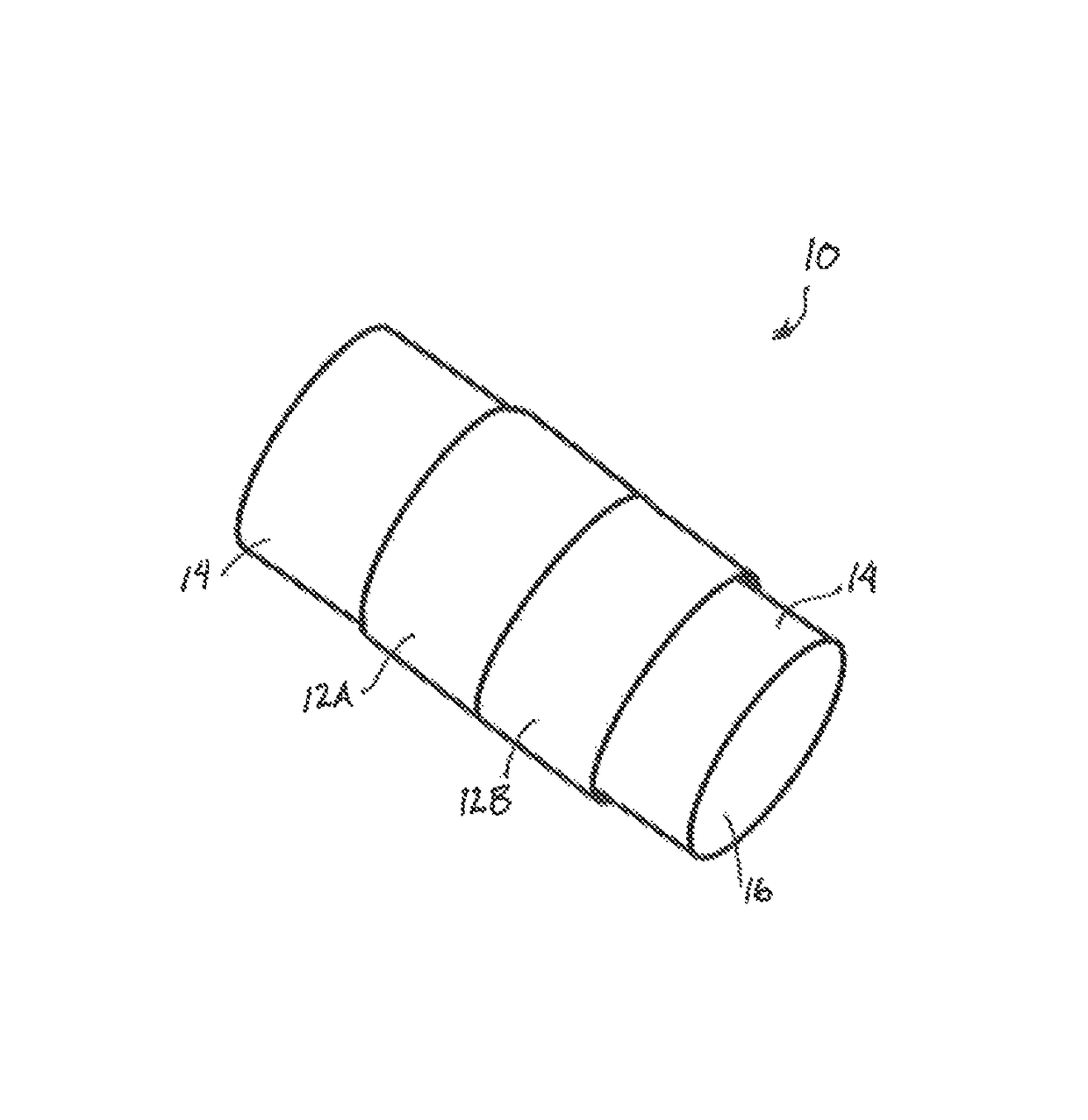
[0082]As shown schematically in FIG. 3, Several circumferentially-oriented circular smooth muscle cell constructs 12A, 12B were bioengineered using a fibrin-based model with an internal diameter of 5 mm. A hollow tubular chitosan scaffold 14 was also manufactured with the following dimensions: 2.5 cm length, 7.25 mm outer diameter, 3.25 mm luminal diameter and 2 mm thickness. The constructs were placed around the scaffold in close proximity. Surgical glue was applied between the junctions of the constructs and along their circumference. The composite tubular tissue structure 10 was maintained in culture for >2 months. The integrity of the muscle tissue was tested by pipetting media through the lumen 16 and flow and leakage were monitored.

[0083]The bioengineered colon segment was 2 cm in length. The construct had a uniform tubular tissue structure similar to native colon. The new bioengineered 2 cm-long circular smooth muscle tubular tissue structure maintained its structural integri...

example 2

[0086]Tubular esophageal tissue was tissue-engineered to mimic the mechanical and physiological function of a native esophagus. Several concentrically aligned esophageal circular smooth muscle tissue constructs were bioengineered using collagen hydrogel. Three-dimensional smooth muscle tissue constructs were bioengineered. Rabbit esophageal constructs were bioengineered by laying down 5×10⁵ rabbit esophageal circular smooth muscle cells in collagen gel on a Sylgard-coated plate with a central post.

[0087]The smooth muscle constructs were placed around a 2.5 cm long hollow chitosan tube and biodegradable surgical glue was applied between the smooth muscle constructs and along their circumference. The constructs were maintained around the scaffold in culture for 5 days.

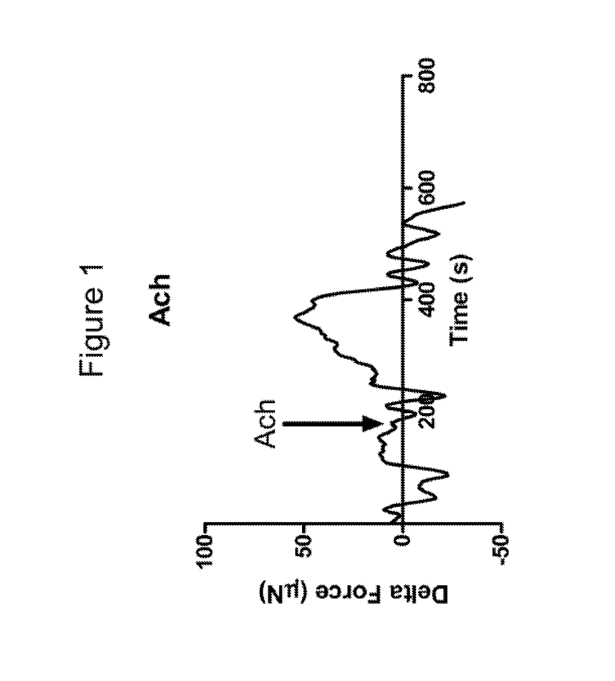
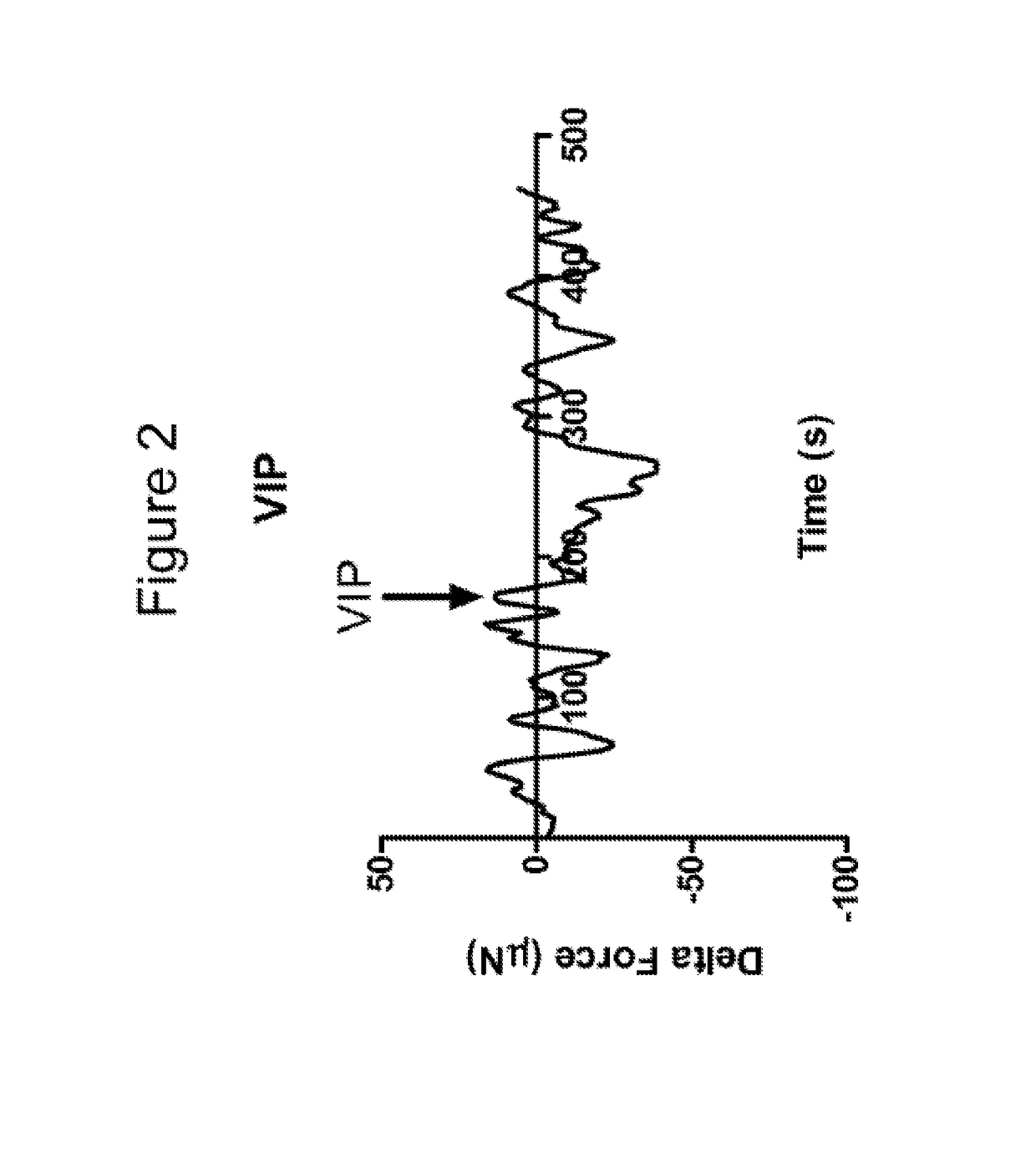
[0088]The constructs were taken off the scaffold and tested to determine cholinergic contraction in response to Acetylcholine (Ach) and relaxation in response to vasoactive intestinal peptide (VIP).

[0089]An isometric for...


PUM

PropertyMeasurementUnit
internal diameteraaaaaaaaaa
thicknessaaaaaaaaaa
luminal diameteraaaaaaaaaa

Abstract

Methods of generating tubular, bioengineered, smooth muscle structures are disclosed as well as bioengineered tissue for tubular organ repair or replacement. The methods can include the steps of obtaining smooth muscle cells; culturing the muscle cells to form a smooth muscle cell construct of directionally oriented smooth muscle cells; disposing the smooth muscle cell construct around a tubular scaffold; and culturing construct and scaffold in a growth media until a smooth muscle cell structure is achieved. The step of obtain smooth muscle cells can further include obtaining autologous smooth muscle cells from a subject. In one preferred embodiment, the muscle cells can first be on a fibrin substrate to form a muscle construct, which is then disposed around a tubular scaffold, for example, a chitosan scaffold. The methods of the present invention can further include connecting two or more tubular structures together to form an elongate composite structure.

Description

REFERENCE TO RELATED APPLICATIONS[0001]This application is based upon and claims the priority of U.S. Provisional Patent Application No. 61 / 592,890, filed Jan. 31, 2012, entitled “Innervation of Engineered Structures, and U.S. Provisional Patent Application No. 61 / 592,871 filed Jan. 31, 2012, entitled “Tubular Bioengineered Smooth Muscle Structures,” which are both hereby incorporated by reference in their entirety.GOVERNMENT RIGHTS[0002]This invention was made with government support under grant NIH / NIDDK RO1DK042876 awarded by The National Institute of Health. The United States Government has certain rights in the invention.FIELD OF THE INVENTION[0003]The present invention concerns tissue engineering of tubular structures, such as gastrointestinal (GI) tissues.BACKGROUND OF THE INVENTION[0004]The human body has numerous tubular structures that perform necessary and vital physiological functions. Smooth muscle cells are the main cell type responsible for proper physiological functi...

Claims


Application Information

Patent Timeline
Patent Type & Authority Patents(United States)
IPC IPC(8): A61K35/34A61L27/20C12N5/00C12N5/077A61L27/38A61L27/50C12N5/0797A61F2/04A61F2/08
CPCA61K35/34A61F2/04A61F2/08A61L27/20A61L27/3826A61L27/3873A61L27/50C12N5/0068C12N5/0623C12N5/0661C08L5/08A61F2002/045A61L2430/30C12N2501/999C12N2533/54C12N2533/72
Inventor BITAR, KHALIL
Owner WAKE FOREST UNIV HEALTH SCI INC
Why Eureka
Try Eureka