
System and method for securing tissue to bone

a tissue and bone technology, applied in the field of medical devices and procedures, can solve the problems of multiple steps and tools, difficult arthroscopic procedures, and problems such as the inability to secure tissue to bone,

Active Publication Date: 2017-11-28
CONMED CORP
View PDF209 Cites 5 Cited by
  • Summary
  • Abstract
  • Description
  • Claims
  • Application Information

AI Technical Summary

Benefits of technology

The patent describes a tissue capture anchor for attaching tissue to bone. The anchor has expandable tines that can be expanded to secure the tissue to the bone. The anchor also includes a spreader that can be inserted into the anchor to deploy the tines. The spreader has a flat tip with holes for sutures to form a loop and capture the tissue. The anchor can be made of polyether-ether-ketone (PEEK) and the tines have teeth to secure the anchor to the bone. The patent also describes a method for inserting the anchor and a clearance hole for the anchor. The technical effect of this invention is to provide a reliable and effective tool for attaching tissue to bone.

Problems solved by technology

Patients with biceps tendon problems may have a detachment of the biceps tendon from the radial tuberosity, for example, or they may have inflammation and irritation of the biceps tendon itself.
Biceps tendon problems can also occur in conjunction with a rotator cuff tear.
This is a difficult procedure arthroscopically.
Systems recently brought to market still require multiple steps and tools.

Method used


Image

  • System and method for securing tissue to bone
  • System and method for securing tissue to bone
  • System and method for securing tissue to bone

Examples


Embodiment Construction

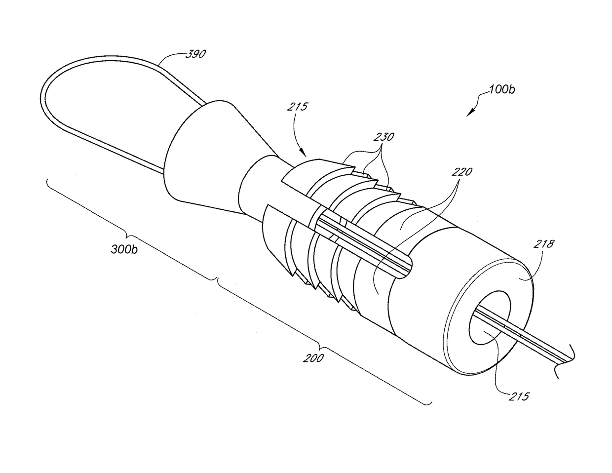
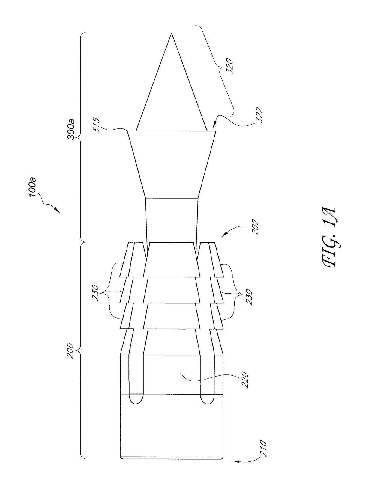
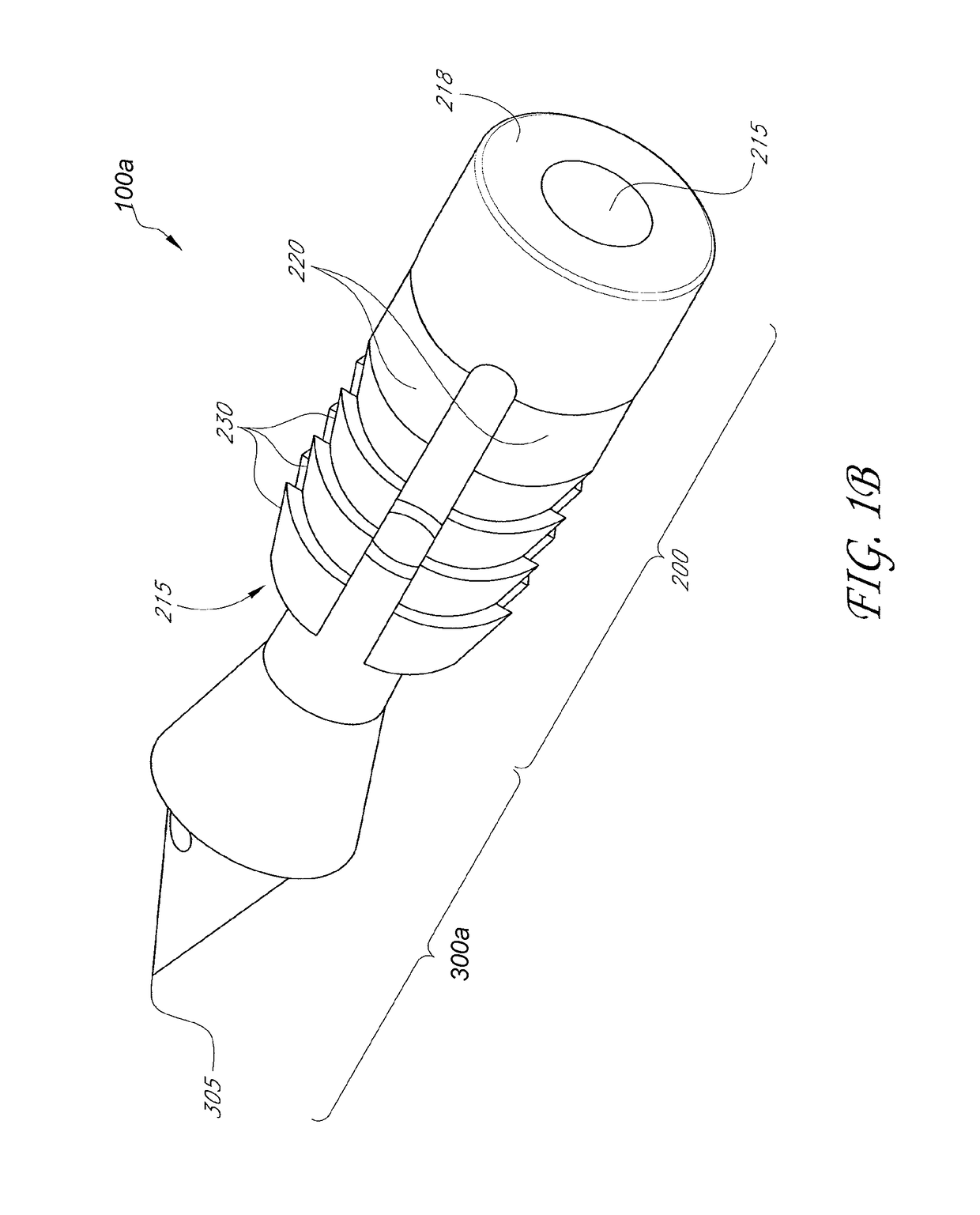
[0072]In various embodiments, soft tissue may be attached to bone utilizing one or more tissue capture anchors. In the following non-limiting examples elements 100a and 100b illustrate two embodiments of a bone anchor, and likewise elements 300a and 300b illustrate a spreader element of the bone anchors. In the following paragraphs, where element 100 is used, it is assumed that elements 100a and 100b are contemplated. Where element 300 is used, it is assumed that elements 300a and 300b are contemplated. The elements 300a and 300b or their corresponding anchor embodiments 100a and 100b are referenced specifically when pertinent.

[0073]In one non-limiting example illustrated in FIGS. 1A-1E, a pointed tip is used to capture tissue. In another non-limiting example illustrated in FIGS. 1F-1J, suture loop is used to capture tissue. FIGS. 1A and 1F depict a side view of a tissue capture anchor 100 comprising an anchor body 200 and a spreader 300. FIG. 1F additionally comprises a suture loop...


PUM


Abstract

Disclosed herein are methods and devices for securing soft tissue to a rigid material such as bone. A tissue capture anchor is described that comprises an anchor body and a spreader such that tissue may be captured or compressed between outside surfaces on the anchor and spreader and inside surfaces of a bone hole to secure the tissue within the hole. A bone anchor is described that comprises an anchor body with expandable tines and a spreader that expands the tines into bone. The spreader captures tissue via a suture loop at the distal end of the bone anchor. Also described is an inserter that can be used to insert the anchor into bone and move the spreader within the anchor to expand the anchor and capture the tissue between the anchor and the bone. Methods are described that allow use of single bone anchor to secure tissue to bone or also to use more than one bone anchor to provide multiple lengths of suture material to compress a large area of soft tissue against bone.

Description

RELATED APPLICATIONS[0001]This application claims priority to U.S. Provisional Application Nos. 61 / 251,220, filed on Oct. 13, 2009; and 61 / 370,791, filed on Aug. 4, 2010; both of which are incorporated herein by reference in their entirety.BACKGROUND OF THE INVENTION[0002]Field of the Invention[0003]The present invention relates to medical devices and procedures. More particularly, the present invention relates to devices and methods for securing soft tissue to a rigid material such as bone.[0004]Description of the Related Art[0005]There are several medical procedures where a surgeon needs to attach soft tissue such as tendons or other soft connective tissue to bone. One common example is a biceps tenodesis, a surgical procedure usually performed for the treatment of biceps tendonitis of the shoulder. A biceps tenodesis may be performed as an isolated procedure, but more often is part of a larger shoulder surgery such as a rotator cuff repair.[0006]The biceps tendon connects the bic...

Claims


Application Information

Patent Timeline
Patent Type & Authority Patents(United States)
IPC IPC(8): A61B17/10A61B17/08A61B17/04
CPCA61B17/0401A61B2017/0409A61B2017/0438A61B2017/0414A61B2017/0424A61B2017/0412A61B17/0483A61B17/0485A61B2017/0432A61B2017/0445A61B2017/0456A61F2/0811A61F2002/0835
Inventor HEAVEN, MALCOLMKO, MICHAELGREELIS, JOHN P.SIRIVONG, MIKXAY
Owner CONMED CORP
Learn More