Diagnostic medical image system and method of introducing Talbot capturing device to diagnostic medical image system used for general capturing

a technology of diagnostic medical image and capturing device, which is applied in the field of diagnostic medical image system, can solve the problems of increasing costs and difficulty in capturing cartilage images

Inactive Publication Date: 2018-01-02
KONICA MINOLTA INC
View PDF18 Cites 16 Cited by
  • Summary
  • Abstract
  • Description
  • Claims
  • Application Information

AI Technical Summary

Benefits of technology

[0047]In the diagnostic medical image system according to the present invention, the controller consolidates several types of diagnostic medical images reconstructed from multiple moire images captured by the second capturing unit into a single data item or group, and sends the consolidated diagnostic medical images to the console, and the console collectively links the several types of diagnostic medical images, which are consolidated into a single data item or group, to a corresponding capturing order information to establish their 1:1 relationship, through a process identical to that of linking the diagnostic medical image captured through a general capturing operation to capturing order information (so as to establish their 1:1 relationship). Alternatively, the controller consolidates image signals corresponding to multiple moire images captured by the second capturing unit into a single data item or group and sends this to the console. The console then reconstructs and generates several types of diagnostic medical images from the image signals corresponding to the multiple moire images.
[0048]This links the several types of diagnostic medical images in the form of a single data item or group to capturing order information. Thus, all of the several types of diagnostic medical images reconstructed from the moire images captured through X-ray capturing performed by the second capturing unit in accordance with the capturing order information can be correctly linked to the capturing order information. This certainly prevents the linking of only one or few of the several types of the diagnostic medical images reconstructed from the moire images captured by the second capturing unit to the capturing order information, which also causes the non-linking of the remaining diagnostic medical images to the capturing order information, or the erroneous determination by the console that other diagnostic medical images remain to be linked to the capturing order information in addition to the diagnostic medical image captured by the first capturing unit, so as to prevent the capturing process from not ending.
[0049]Data on one diagnostic medical image captured by the first capturing unit, data on multiple moire images captured by the Talbot capturing device, and data on the several types of diagnostic medical images reconstructed and generated from the moire images coincide in the system. Thus, the data on the diagnostic medical image can be prevented from being erroneously linked to unrelated capturing order information, or a diagnostic medical image captured by a general-purpose capturing unit can be certainly prevented from being erroneously linked to unrelated capturing order information that is already linked to several types of diagnostic medical images.

Problems solved by technology

It is difficult to capture an image of the cartilage portion in a joint region of a patient of interest in general capturing.
Such capturing involving MRI applies physical and mental stress to the patient who is the subject and causes an increase in costs.

Method used


Image

  • Diagnostic medical image system and method of introducing Talbot capturing device to diagnostic medical image system used for general capturing
  • Diagnostic medical image system and method of introducing Talbot capturing device to diagnostic medical image system used for general capturing
  • Diagnostic medical image system and method of introducing Talbot capturing device to diagnostic medical image system used for general capturing

Examples


first embodiment

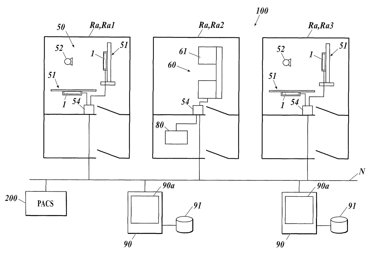
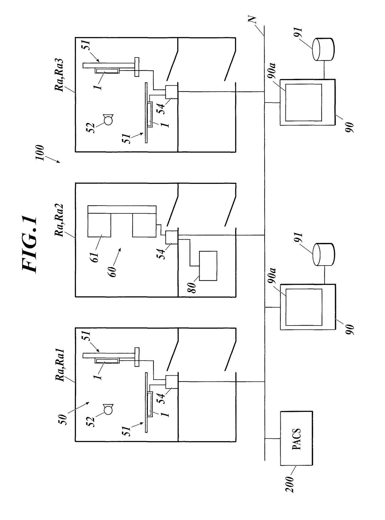
[0073]FIG. 1 illustrates an example configuration of a diagnostic medical image system according to a first embodiment of the present invention. In this embodiment, a diagnostic medical image system 100 includes a plurality of capturing rooms Ra (Ra1 to Ra3) and a plurality of consoles 90. Components in the capturing rooms Ra are connected to the consoles 90 via a network N. In this embodiment, the diagnostic medical image system 100 is connected to a PACS 200 via the network N, and also to HISs and RISs via the network N, although not shown in the drawings.

[0074]In the diagnostic medical image system 100 illustrated in FIG. 1, capturing rooms Ra containing first capturing units 50 (i.e., capturing rooms Ra1 and Ra3) do not contain second capturing units 60, whereas a capturing room Ra containing a second capturing unit 60 (i.e., capturing room Ra2) does not contain a first capturing unit 50. Alternatively, a single capturing room Ra may contain both a first capturing unit 50 and a ...

modification 1

[Modification 1]

[0140]Consoles 90 and PACSs 200 included in conventional diagnostic medical image systems for general capturing are mostly configured to manage and display diagnostic medical images (and combined images if present (the same applies hereinafter)) having a normal gradation, e.g., a 12-bit gradation. Diagnostic terminals and dedicated terminals have been developed that can display diagnostic medical images having a high gradation of 16 bits, for example. Thus, the controller 80 may select between two modes for reconstruction and generation of several types of diagnostic medical images: a normal mode for generating a diagnostic medical image having a normal gradation, e.g., a 12-bit gradation, and a high-gradation mode for generating images in a higher gradation.

modification 1-1

[Modification 1-1]

[0141]If the console 90 can process a normal 12-bit gradation, the controller 80 carries out WW / WC gradation processing to the diagnostic medical image generated in a normal 12-bit mode; DICOM processing is carried out to both diagnostic medical images generated in the normal mode and the high-gradation mode; and the diagnostic medical images generated in the normal mode and the diagnostic medical images generated in the high-gradation mode are respectively consolidated and sent to the console 90. As described above, the console 90 is operated to consolidate the several types of diagnostic medical images to be linked with capturing order information. The capturing order information can be duplicated, and the diagnostic medical image generated in the normal mode may be linked to one of the capturing order information and the diagnostic medical images generated in the high-gradation mode to the other capturing order information. In such a case, the two pieces of capt...


PUM


Abstract

Provided are a diagnostic medical image system and the like which enable diagnostic medical images generated in either an imaging system used for general imaging or an imaging system which includes a Talbot imaging device to be accurately associated with imaging order information. In the diagnostic medical image system, a controller groups a plurality of types of diagnostic medical images reconstructed and generated on the basis of a plurality of image signals captured by a second imaging system equipped with a Talbot imaging device and transmits the group to a console; and the console associates one diagnostic medical image generated from one image signal acquired from a first imaging system equipped with an X-ray imaging device with the imaging order information corresponding thereto and, by the same associating method, associates the grouped plurality of types of diagnostic medical images transmitted from the controller with the imaging order information corresponding thereto.

Description

CROSS REFERENCE TO RELATED APPLICATIONS[0001]This is the U.S. national stage of application No. PCT / JP2014 / 054514, filed on Feb. 25, 2014. Priority under 35 U.S.C. §119(a) and 35 U.S.C. §365(b) is claimed from Japanese Application No. 2013-080273, filed Apr. 8, 2013, and from Application No. 2013-0256561, filed Dec. 12, 2013, the disclosures of which are also incorporated herein by reference.TECHNICAL FIELD[0002]The present invention relates to a diagnostic medical image system including a first capturing unit for performing general capturing with an X-ray source and an X-ray capturing device, and a second capturing unit for capturing several types of diagnostic medical images with a Talbot capturing device.BACKGROUND ART[0003]In the field of X-ray capturing, the transition of X-ray capturing devices from conventional silver halide photographic devices including screens or films to CR (computed radiographic) units including photostimulable phosphor sheets has led to transition to X-...

Claims


Application Information

Patent Timeline
Patent Type & Authority Patents(United States)
IPC IPC(8): A61B6/00A61B6/04
CPCA61B6/566A61B6/4233A61B6/4266A61B6/4283A61B6/4417A61B6/463A61B6/483A61B6/484A61B6/5235A61B6/5247A61B6/563A61B6/4035A61B6/467A61B6/0407A61B6/4452A61B6/465
Inventor HAMANO, MASAYAKURAHASHI, AKIRAMATSUSAKA, NORIHIROKIYOHARA, JUNKOYOKOYAMA, TOMOYASUNISHINO, SATOSHI
Owner KONICA MINOLTA INC
Why Eureka
Try Eureka