Body fluid analysis system as well as image processing device and method for body fluid analysis

A technique of body fluids, images

Inactive Publication Date: 2011-05-11
SIEMENS AG
View PDF8 Cites 8 Cited by
  • Summary
  • Abstract
  • Description
  • Claims
  • Application Information

AI Technical Summary

Problems solved by technology

However, mechanical adjustment is less reliable

Method used


Image

  • Body fluid analysis system as well as image processing device and method for body fluid analysis
  • Body fluid analysis system as well as image processing device and method for body fluid analysis
  • Body fluid analysis system as well as image processing device and method for body fluid analysis

Examples


Embodiment Construction

[0072] In order to make the objectives, technical solutions, and advantages of the present invention clearer, the following further describes the present invention in detail with reference to the accompanying drawings and embodiments.

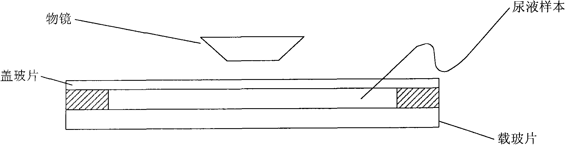
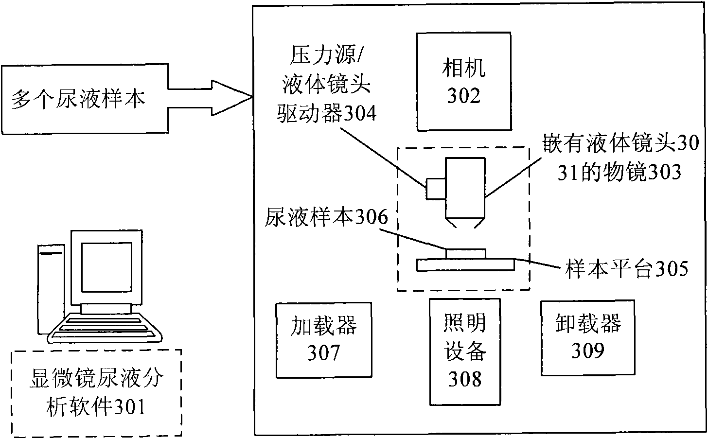
[0073] Figure 3(a) is a schematic diagram of the composition of an image-based body fluid analysis system in an embodiment of the present invention. The following only takes urine as an example to illustrate the working principle of the body fluid analysis system. Of course, the system is also suitable for the analysis of blood, cerebrospinal fluid, pleural effusion, ascites, semen and other body fluids.

[0074] In the system shown in FIG. 3(a), the microscope urine analysis software 301 is used to analyze and process the images taken by the camera 302. An objective lens 303 embedded with a liquid lens 3031 is connected to the camera 302, and the objective lens 303 also includes a zoom lens 3032. The pressure source / liquid lens driver 304 is used ...


PUM


Abstract

The invention discloses a body fluid analysis system, comprising a central control and process unit and a source image capture unit, wherein the central control and process unit is used for sending a control signal to the source image capture unit; the source image capture unit is used for capturing a body fluid source image according to the control signal, and sending the source image to the central control and process unit; and the central control and process unit is further used for transforming the source image to an image coefficient, generating a corresponding coefficient matrix, and then inverse-converting the coefficient matrix to an image output after a focus fusion. The invention further discloses an image processing device and method for body fluid analysis. According to the invention, the probability of missing or blurring of the target image can be reduced, thereby significantly enhancing the success rate of the recognition and improving the accuracy of the whole system.

Description

Technical field [0001] The invention relates to the field of body fluid analysis, and in particular to an image processing device and method used in an image-based automatic microscope body fluid analyzer, and a body fluid analysis system. Background technique [0002] Body fluid analysis, especially microscopic urine sediment analysis, is one of the most commonly performed tests in clinical practice because it can provide critical information for kidney and genitourinary diagnosis, as well as overall health. More than 10 types of particles can be found in urine samples, including: red blood cells, white blood cells, sloughs, bacteria, epithelial cells, and crystals (hereinafter these particles are referred to as objects). These particles with different shapes and sizes need to be identified and counted to generate a spectrum reflecting the proportions of different types of elements, which can be compared with thresholds or health reference values. [0003] In terms of urine sedim...

Claims


Application Information

Patent Timeline
Patent Type & Authority Applications(China)
IPC IPC(8): G01N15/04
CPCG01N15/1434G01N15/1429G01N15/1463G01N2015/1452G01N2015/1477G02B21/367G02B26/004G06T5/003G06T5/50G06T2207/10016G06T2207/10056G06T2207/20221G06T2207/30004
Inventor 董潇潇杜昭辉
Owner SIEMENS AG
Why Eureka
Try Eureka