Small animal living body multi-mode molecule imaging system and imaging method

A molecular imaging and small animal technology, applied in the field of image processing, can solve problems such as weak measurement signal, excessive computational complexity, and difficulty in solving high-order approximate equations

Active Publication Date: 2012-06-13
XIDIAN UNIV
View PDF6 Cites 56 Cited by
  • Summary
  • Abstract
  • Description
  • Claims
  • Application Information

AI Technical Summary

Problems solved by technology

Higher-order approximations to the radiative transfer equation enable accurate imaging of small animal objects with both high-scattering and low-scattering tissue, see: J.Zhong, J.Tian, ​​X.Yang, C.Qin, Whole-Body Cerenkov Luminescence Tomography with the Finite Element SP3 Method, Annals of Biomedical Engineering, 39: 1728-1735 (2011). This method can guarantee a certain solution accuracy, but it is very difficult to solve high-order approximation equations. For inhomogeneous light with com

Method used


Image

  • Small animal living body multi-mode molecule imaging system and imaging method
  • Small animal living body multi-mode molecule imaging system and imaging method
  • Small animal living body multi-mode molecule imaging system and imaging method

Examples


Embodiment Construction

[0058] Various details involved in the technical solution of the present invention will be described in detail below in conjunction with the accompanying drawings. The described examples are only intended to facilitate the understanding of the invention.

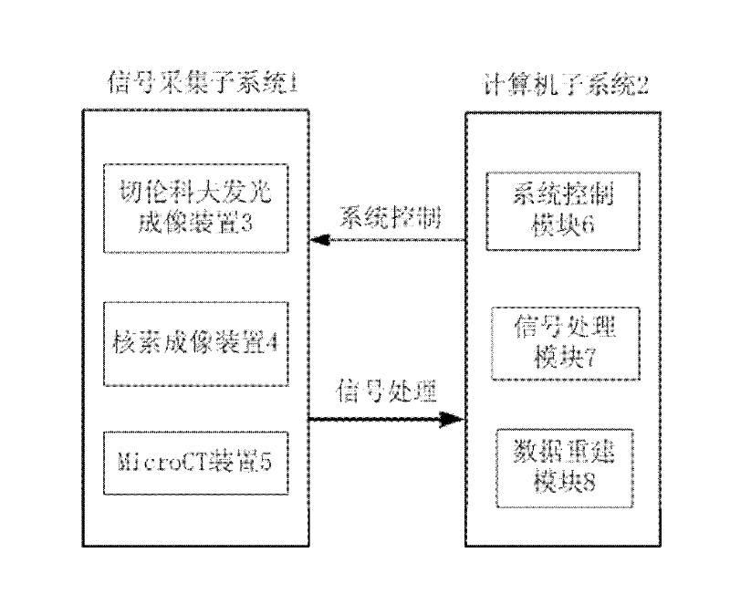
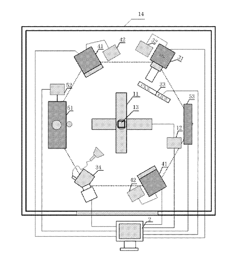
[0059] refer to figure 1 and figure 2 , the small animal in vivo multi-modal molecular imaging system of the present invention includes: a signal acquisition subsystem 1 and a computer subsystem 2, wherein:

[0060] The signal acquisition subsystem 1 includes a Cerenkov luminescence imaging device 3 , a nuclide imaging device 4 and a MicroCT device 5 , a rotary translation stage 11 , an electric translation and rotation stage control box 12 , a small animal support 13 and a dark box 14 . The Cerenkov luminescent imaging device 3 includes a CCD detector 31, a CCD detector power box 32, a filter set 33 and a scanning spectrometer device 34; the nuclide imaging device 4 includes two PET detection 41 and a detector The power s...


PUM


Abstract

The invention discloses a small animal living body multi-mode molecule imaging system and an imaging method. The imaging system comprises a Cerenkov luminous imaging device, a nuclide imaging device and a MicroCT (micro computed tomography) device which respectively acquire Cerenkov optical signals, nuclide signals and X-ray signals of body surfaces of small animals. The X-ray signals are reconstructed by the aid of cone-beam back-projection algorithm, and information of an anatomical structure of a small animal is obtained; the Cerenkov optical signals are reconstructed on the basis of a mixed light transmission theory, and three-dimensional distribution information of a Cerenkov target is obtained; and the obtained Cerenkov target information and the obtained anatomical structure information are fused and displayed, so that multi-mode imaging is completed. A single-mark probe/multi-mode imaging mode which integrates high specificity of nuclide imaging, high sensitivity of optical imaging and high resolution of X-ray imaging is realized, so that multi-mode image information of small animal living bodies is more complete, comprehensive and accurate.

Description

technical field [0001] The invention belongs to the technical field of image processing, and relates to nuclide imaging and Cherenkov luminescence imaging based on radionuclide, MicroCT imaging, especially a multi-modal molecular imaging device and an optical imaging three-dimensional imaging method, which can be used for in vivo molecules of small animals imaging. Background technique [0002] Molecular imaging technology has achieved rapid development in the past decade, and has been widely used in small animal experiments and pre-clinical experiments such as tumor detection and drug development. At present, the commonly used imaging technologies mainly include nuclide imaging, MRI, and optical imaging. These modalities have different characteristics, and have their own advantages and disadvantages in terms of system specificity, sensitivity, and equipment cost. Nuclide imaging, such as PET and SPECT imaging, has high sensitivity and is not limited by the detection depth,...

Claims


Application Information

Patent Timeline
IPC IPC(8): A61B5/00A61B6/03
CPCA61B6/508
Inventor 屈晓超梁继民赵凤军陈雪利朱守平陈多芳刘俊廷胡振华田捷
Owner XIDIAN UNIV
Why Eureka
Try Eureka