
Endoscopic image lesion detection method based on fusion of global and local features

A technology of local features and global features, applied to computer parts, character and pattern recognition, instruments, etc., can solve the problems of time and energy for professional doctors, incomplete labeling of local lesion areas, and affecting the accuracy of lesion detection, etc., to achieve Reduce the labor intensity of doctors, ensure the accuracy of lesion detection, and reduce the effect of missed detection rate

Inactive Publication Date: 2012-10-10
SOUTHWEST JIAOTONG UNIV
View PDF0 Cites 34 Cited by
  • Summary
  • Abstract
  • Description
  • Claims
  • Application Information

AI Technical Summary

Problems solved by technology

This makes it necessary for professional doctors to spend a lot of time and energy on labeling training images when extracting local features to train the classifier, and it is prone to incomplete or wrong labeling of local lesion areas, which affects the accuracy of lesion detection

Method used


Image

  • Endoscopic image lesion detection method based on fusion of global and local features
  • Endoscopic image lesion detection method based on fusion of global and local features
  • Endoscopic image lesion detection method based on fusion of global and local features

Examples


Embodiment Construction

[0038] The following examples are only used to illustrate the present invention, but are not intended to limit the application scope of the present invention:

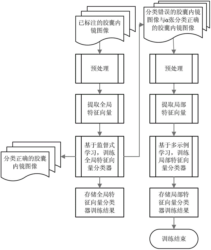
[0039] A. Construct a capsule endoscopy image training sample library with labeled information, extract the global and local features of the capsule endoscopy small intestine image, and train a capsule endoscopy image classifier.

[0040] A.1 Extract the global features of the capsule endoscopic image, and train the global feature classifier.

[0041] 1) Use various computer means and techniques to collect images of capsule endoscopy of patients. Classify and sort endoscopic images by professional doctors, and mark whether there are lesions in the images; mark the lesion types for images with lesions (no need to mark the specific position of the lesion area in the image), and establish an in-capsule capsule with marked information Endoscopic image training sample library; in this embodiment, we use 2000 capsule endosc...


PUM


Abstract

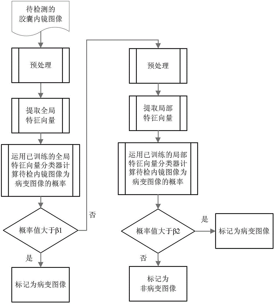
The invention discloses an endoscopic image lesion detection method based on based on fusion of global and local features. According to the method, supervised learning and multi-instance learning are combined, two levels of lesion detection mechanisms are established, the rate of missing detection is farthest reduced under the condition that the lesion detection accuracy and reducing labor intensity of doctors are ensured at the same time, and missed diagnosis or error diagnosis due to that a lesion image is mistakenly identified to a non-lesion image is avoided. According to the endoscopic image lesion detection method based on based on fusion of global and local features, only whether a trained image has lesion or not needs to be labeled and a specific lesion region does not need to be labeled when local features are extracted for training a classifier due to introduction of the multi-instance learning, thus the method is more convenient and easy in practical application than conventional method.

Description

Technical field [0001] The invention belongs to digital image processing technology, in particular the combination of machine learning technology and clinical capsule endoscope small intestine image lesion detection. Background technique [0002] Capsule endoscopy is a new type of gastrointestinal disease detection tool, which has the advantages of painlessness, safety, and full detection. It has been widely used in clinical practice at home and abroad, and achieved good results. Usually, a capsule endoscopy test will produce 40,000 to 60,000 color images, which are screened and analyzed by professional doctors to diagnose various gastrointestinal lesions. However, it takes 1 to 2 hours for a professional doctor just to browse the images. Therefore, it is urgent to establish a computer-aided system for lesion detection in capsule endoscopic images, exclude non-lesion images and strong interference images, and submit the screened images of suspected lesions to doctors for fu...

Claims


Application Information

Patent Timeline
IPC IPC(8): G06K9/66
Inventor 陈俊周李青张理彭强
Owner SOUTHWEST JIAOTONG UNIV
Learn More