AI technical title is built by Patsnap AI team. It summarizes the technical point description of the patent document.
A detection method and CT image technology, applied in image data processing, instrumentation, calculation, etc., can solve problems such as high system performance and time consumption, and low efficiency of lesion detection methods
Inactive Publication Date: 2019-01-08
NEUSOFT MEDICAL SYST CO LTD
View PDF0 Cites 12 Cited by
Summary
Abstract
Description
Claims
Application Information
AI Technical Summary
This helps you quickly interpret patents by identifying the three key elements:
Problems solved by technology
Method used
Benefits of technology
Problems solved by technology
[0005] Since the existing processing method requires two convolution calculations, which contain repeated calculation content, and the convolution calculation itself is a processing process that consumes a lot of system performance and time, the current lesion detection method is inefficient.
Method used
Image
Click on the blue label to locate the original text in one second.
Reading with bidirectional positioning of images and text.
Examples
Embodiment 1
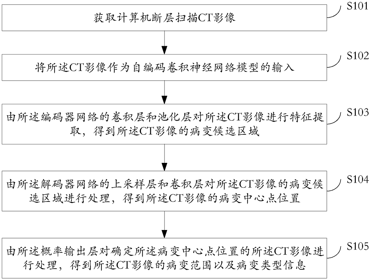
[0049] see figure 1 , which is a flow chart of a lesion detection method provided in an embodiment of the present application, the method comprising:
[0051] In the embodiment of the present application, before lesion detection, first obtain a CT image as a lesion detection object; wherein, the CT image can be a CT image of the patient's organ to be detected, such as a lung CT image, a thyroid CT image, a brain CT image, etc. Images, etc. Specifically, the embodiment of the present application can detect pulmonary nodules in lung CT images, detect thyroid nodules in thyroid CT images, and detect cerebral ischemia and necrosis in brain CT images. etc., and no specific lesion detection object is limited here.
[0052] In practical applications, the CT image used as the lesion detection object may be a CT image acquired in advance and stored in a database, or may be a CT image acquired after performing a CT scan on a patien...
Embodiment 2
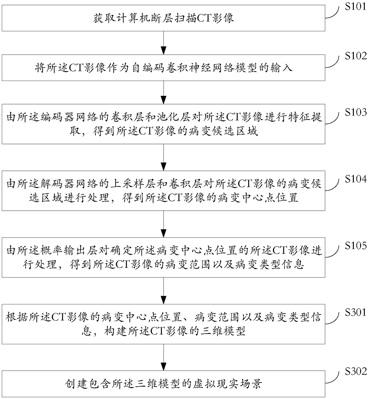
[0071] For the location of the lesion center point, lesion range, and lesion type information detected after lesion detection, doctors need to further diagnose the patient based on the above test results. In order to facilitate doctors to understand the patient's test results more intuitively, and To determine the follow-up treatment more accurately, refer to image 3 , the embodiment of the present application further provides the following steps on the basis of the above method embodiment 1 S101-S105:
[0072] S301: Construct a three-dimensional model of the CT image according to the lesion center position, lesion range, and lesion type information of the CT image.
[0073] After detecting the lesion center point position, lesion range and lesion type information of the CT image through the first embodiment of the above method, a three-dimensional model of the CT image is constructed based on the above detection results.
[0074] Taking the construction of the 3D model of t...
PUM
Abstract
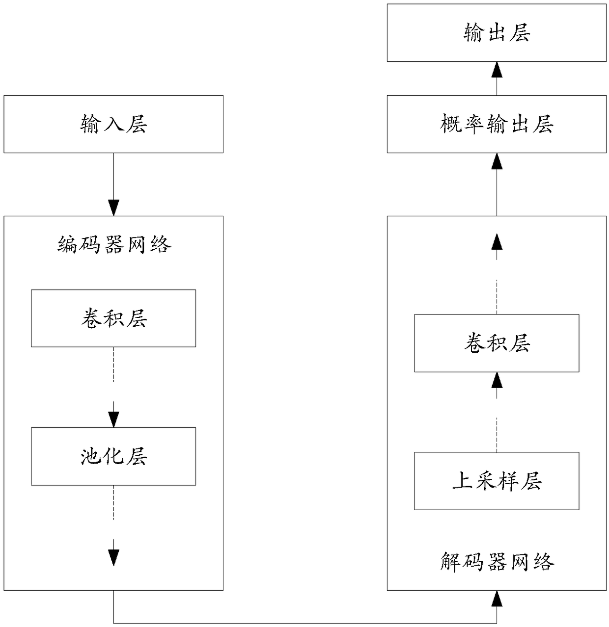
A lesion detection method, apparatus and device are disclosed. The method comprises: a CT image is processed by a self-encodeconvolutional neural network model, feature extraction is conducted on theCT image by a convolution layer and a pooling layer of the encoder network, a lesion candidate region of the CT image is obtained, the lesion candidate region of CT image is processed by the upper sampling layer and convolution layer of the decoder network, and the lesion center position of the CT image is obtained; the CT image which has determined the lesion center position is processed by theprobability output layer, and the lesion range and the lesion type information of the CT image are obtained. Since the calculation of the decoder network is based on the convolutionprocessing resultof the encoder network, and the application does not need to carry out repeated convolution calculation processing, thus saving the convolution calculation processing time and improving the lesion detection efficiency to a certain extent on the premise of ensuring the lesion detection accuracy.
Description
technical field [0001] The present application relates to the field of medical image processing, in particular to a lesion detection method, device and equipment. Background technique [0002] Cancer is currently the most threatening disease to human health and life in society. Its commonly used diagnostic technology is computerized tomography (CT) technology, which is usually used to diagnose whether various organs have lesions and determine the type of lesions ( That is, benign and malignant), it is helpful for the early diagnosis and treatment of various cancers, and thus can reduce the mortality of patients. [0003] For example, lungcancer is one of the fastest-growing cancers in morbidity and mortality. Lungcancer generally evolves from pulmonary nodules. Doctors can help early diagnosis and treatment of lung cancer by analyzing the benign and malignant of lung nodules in patients. [0004] In actual clinical application, the full convolutional neural network model ...
Claims
Application Information
Patent Timeline
Application Date:The date an application was filed.
Publication Date:The date a patent or application was officially published.
First Publication Date:The earliest publication date of a patent with the same application number.
Issue Date:Publication date of the patent grant document.
PCT Entry Date:The Entry date of PCT National Phase.
Estimated Expiry Date:The statutory expiry date of a patent right according to the Patent Law, and it is the longest term of protection that the patent right can achieve without the termination of the patent right due to other reasons(Term extension factor has been taken into account ).
Invalid Date:Actual expiry date is based on effective date or publication date of legal transaction data of invalid patent.

