Acetabular tissue model reconstruction method for serialized hip joint CT images
A technology of CT images and hip joints, applied in image data processing, 3D modeling, instruments, etc., can solve problems such as large differences in segmentation results, complex manual interaction, and difficulty in accumulating samples, and achieve easy programming and algorithm complexity Low, easy-to-operate effect
- Summary
- Abstract
- Description
- Claims
- Application Information
AI Technical Summary
Problems solved by technology
Method used
Image
Examples
Embodiment 1
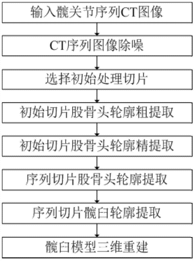
[0063] Example 1, such as figure 1 A method for reconstructing an acetabular tissue model oriented to serialized hip joint CT images as shown, comprising the following steps:
[0064] CT image preprocessing: 3D Gaussian blur method is used to denoise each CT slice in the CT image sequence.
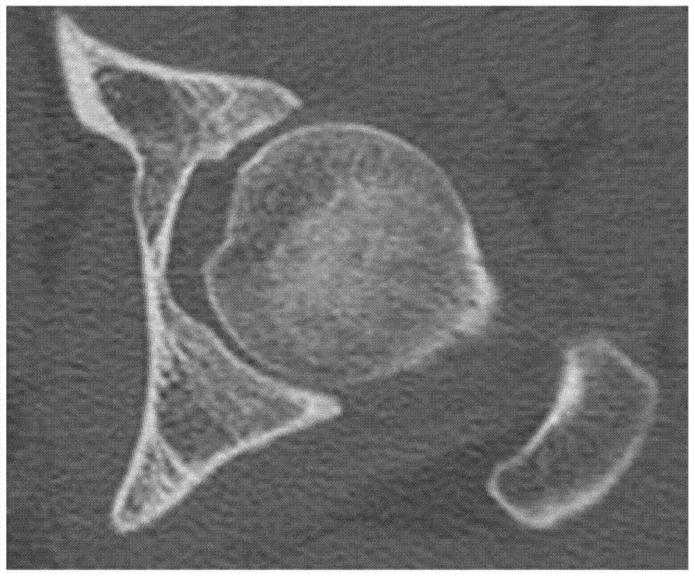
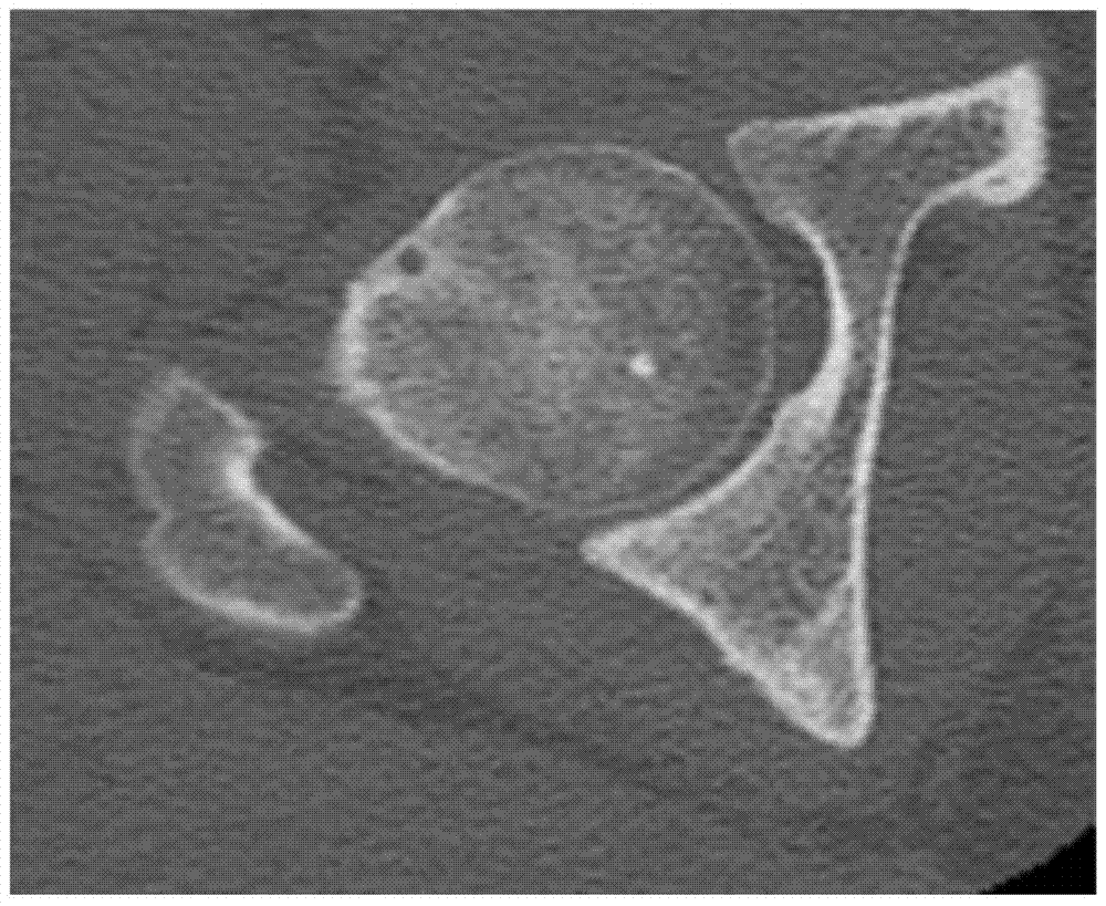
[0065] Select the initial processing slice: the CT slice where the greater trochanter and femoral head were separated for the first time was used as the initial CT slice. Such as figure 2 Shown is an image slice of the left hip joint, image 3 Shown is an image slice of the right hip.
[0066] Create a space Cartesian coordinate system:
[0067] Take the upper left corner of the first slice in the CT image sequence as the coordinate origin, take the rightward direction as the positive direction of the x-axis, take the downward direction as the positive direction of the y-axis, and take the direction in which the number of slice layers increases as the positive direction of the z-axis ...
PUM
 Abstract
Description
Claims
Application Information
 - R&D
- Intellectual Property
- Life Sciences
- Materials
- Tech Scout
- Unparalleled Data Quality
- Higher Quality Content
- 60% Fewer Hallucinations
Browse by: Latest US Patents, China's latest patents, Technical Efficacy Thesaurus, Application Domain, Technology Topic, Popular Technical Reports.
© 2025 PatSnap. All rights reserved.Legal|Privacy policy|Modern Slavery Act Transparency Statement|Sitemap|About US| Contact US: help@patsnap.com


