Clinical workstation integrating medical imaging and biopsy data and methods using same

A workstation, medical image technology, used in special data processing applications, medical equipment, medical images, etc., can solve problems such as the integration of non-medical imaging workflow and biopsy workflow

Active Publication Date: 2015-01-21
KONINKLJIJKE PHILIPS NV
View PDF6 Cites 11 Cited by
  • Summary
  • Abstract
  • Description
  • Claims
  • Application Information

AI Technical Summary

Problems solved by technology

[0007] However, existing medical solutions do not effe

Method used


Image

  • Clinical workstation integrating medical imaging and biopsy data and methods using same
  • Clinical workstation integrating medical imaging and biopsy data and methods using same
  • Clinical workstation integrating medical imaging and biopsy data and methods using same

Examples


Embodiment Construction

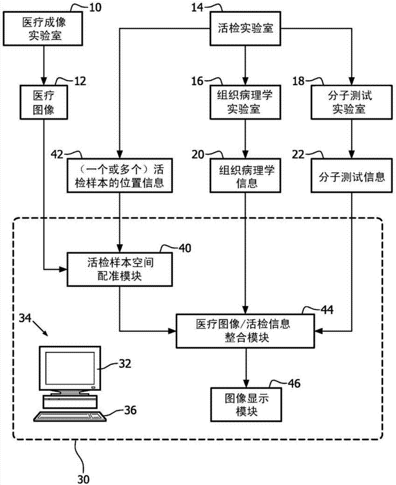
[0029] refer to figure 1 , the medical imaging laboratory 10 uses an appropriate imaging modality such as magnetic resonance (MR) imaging, positron emission tomography (PET) imaging, single photon emission computed tomography (SPECT), transmission computed tomography (CT), or Other transmission X-ray imaging techniques and the like acquire medical images 12 of medical objects. The medical image 12 may be two-dimensional, three-dimensional or four-dimensional (eg, a cine or CINE sequence tracking intake and / or washout of a bolus injection).

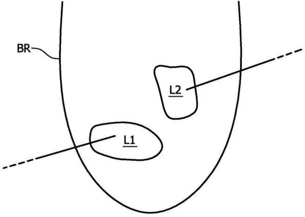
[0030] As used herein, the term "medical subject" includes hospital patients, out-of-hospital patients (eg, patients who are not admitted to hospital but receive medical care), clinical research subjects, medical screening subjects, veterinary subjects, and the like. The medical image 12 is designed to elicit relevant information about the medical subject's medical condition. In the illustrative case of oncology medical subjects, medical...


PUM


Abstract

An imaging visualization workstation (30) includes a graphical display device (32) and an electronic data processor, and is configured to perform a method including: spatially registering a biopsy sample extracted from a medical subject with a medical image (12) of the medical subject; combining the medical image with a graphical representation of information (20, 22) generated from the biopsy sample to generate a combined image in which the graphical representation is spatially delineated based on the spatial registration of the biopsy sample; and displaying the combined image on the graphical display device of the imaging visualization workstation. A method comprises extracting a biopsy sample from a medical subject, processing the biopsy sample to generate biopsy information, acquiring a medical image of the subject, spatially registering the biopsy sample with the medical image, and displaying the medical image modified to include an annotation generated from the biopsy information.

Description

technical field [0001] The following relates to medical technology, oncology technology, medical imaging technology, biopsy and pathology technology, and related technologies. Background technique [0002] Medical imaging is used in oncology and other medical fields to provide noninvasive or minimally invasive characterization of the structure and / or function of cancerous tumors, organs containing metastatic cancer cells, or other anatomical structures of interest. Various medical imaging modalities are known and their diverse imaging capabilities can be exploited. Some of the imaging modalities used in various types of medical imaging include: Magnetic resonance (MR) imaging; Positron emission tomography (PET) imaging; Single photon emission computed tomography (SPECT); Transmission computed tomography (CT) or other transmission X-ray imaging techniques; etc. Some examples of imaging techniques include: static anatomical imaging (with or without exogenous contrast agent);...

Claims


Application Information

Patent Timeline
IPC IPC(8): G06F19/00A61B6/00G16H10/60G16H30/20G16H30/40G16H40/60
CPCG16H10/40G16H30/40G16H30/20G16H40/60
Inventor N·班纳吉S·卡玛拉卡兰V·瓦拉达恩A·亚内夫斯基N·迪米特罗娃
Owner KONINKLJIJKE PHILIPS NV
Why Eureka
Try Eureka