Eureka AIR delivers breakthrough ideas for toughest innovation challenges, trusted by R&D personnel around the world.

perfusion imaging

An imaging method, 4D technology, applied in image analysis, image enhancement, image generation, etc., can solve the problem of not providing comprehensive analysis of heterogeneous tissue perfusion parameters

Active Publication Date: 2018-01-26
KONINKLJIJKE PHILIPS NV
View PDF10 Cites 0 Cited by
  • Summary
  • Abstract
  • Description
  • Claims
  • Application Information

AI Technical Summary

Problems solved by technology

Unfortunately, such methods still do not provide comprehensive analysis of perfusion parameters in heterogeneous tissues

Method used


Image

  • perfusion imaging
  • perfusion imaging
  • perfusion imaging

Examples


Embodiment Construction

[0027] A method for analyzing the perfusion properties of spatially entangled heterogeneous tissue in 4D image data is described below, in which spatially entangled tissue components are separated and generated and visually presented Perfusion maps for individual tissue components.

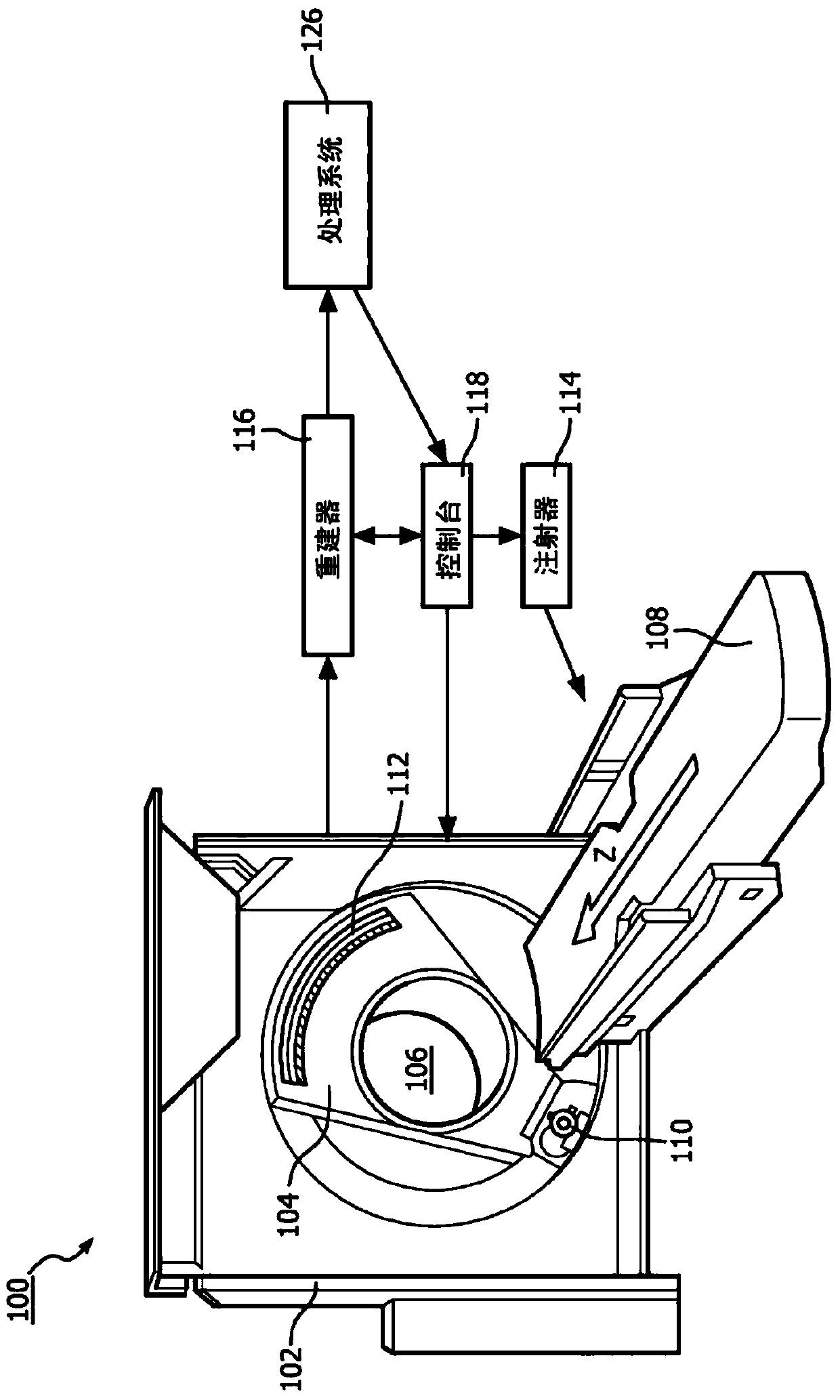
[0028] figure 1 An example imaging system 100 such as a CT scanner is schematically illustrated. In other embodiments, imaging system 100 may include one or more of: MRI, PET, SPECT, US, combinations thereof, and / or other imaging systems configured to perform dynamic contrast-enhanced imaging scans. The illustrated imaging system 100 includes a fixed gantry 102 and a rotating gantry 104 that is rotatably supported by the fixed gantry 102 and rotates about an examination region 106 about a z-axis.

[0029] A radiation source 110 , such as an X-ray tube, is rotatably supported by the rotating gantry 104 , rotates with the rotating gantry 104 , and emits polychromatic radiation through the examinat...


PUM


Abstract

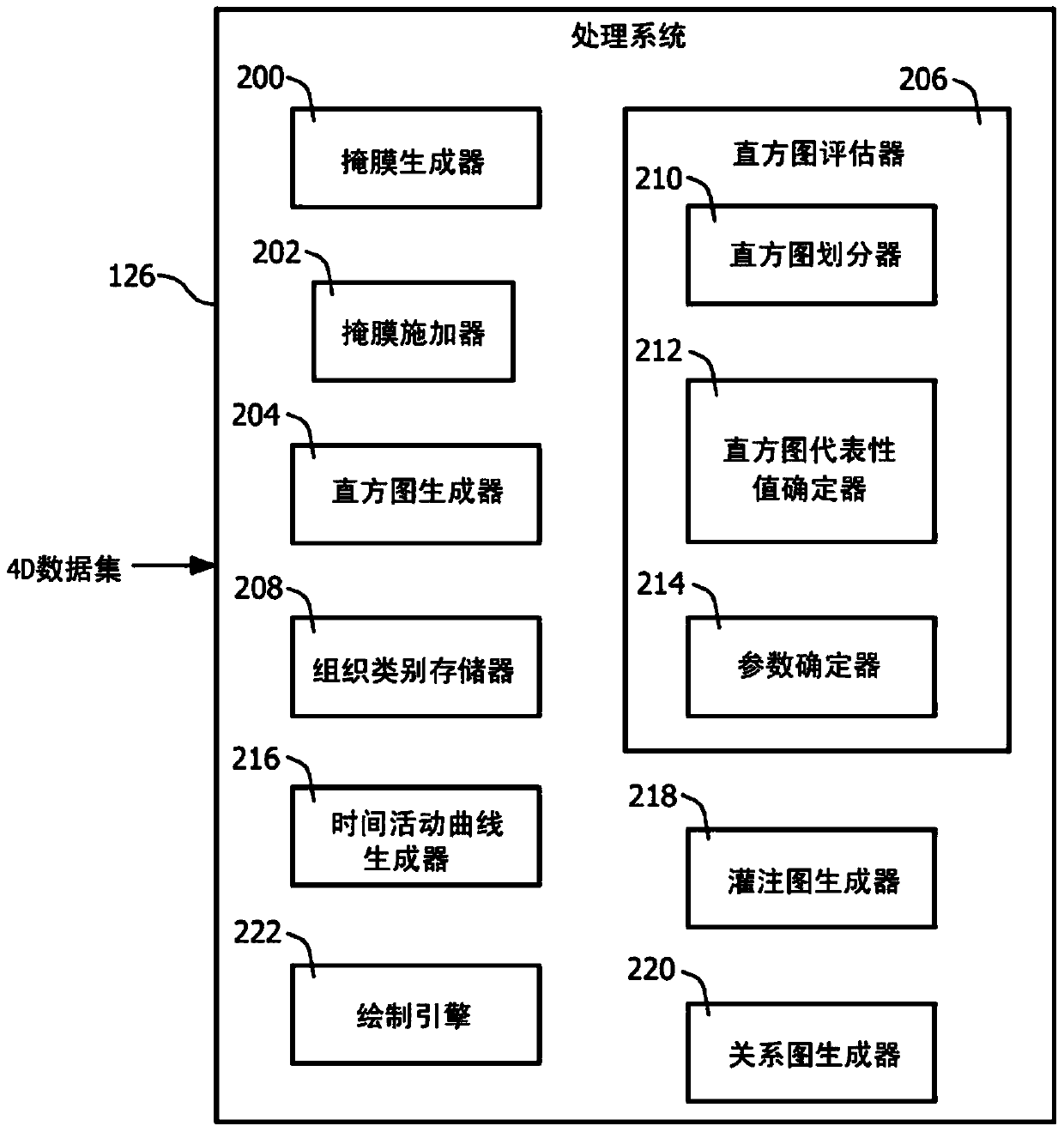
Described herein is a method for analyzing the perfusion properties of heterogeneous tissues in a 4D image dataset (i.e., a time series of contrast-enhanced 3D volumes), in which spatially entangled tissue components are separated into individual and generate and visually present a perfusion map for the individual's tissue composition. In one example, the method comprises: obtaining the 4D dataset in electronic format, generating for each time frame being assessed a time frame for each of the different tissue components for each voxel being assessed different time-activity curve points, and generating a signal indicative of a different parametric map for each of the different tissue components based at least on the time-activity curve. Optionally, relationships between parameters of different components are determined and presented in a relationship graph.

Description

technical field [0001] The following generally refers to perfusion imaging and is described with specific application to computed tomography (CT); however, the following also applies to other imaging modalities, including magnetic resonance imaging (MRI), positron Emission Tomography (PET), Single Photon Emission Computed Tomography (SPECT), Ultrasound (US), and / or other imaging modalities. Background technique [0002] CT perfusion imaging uses contrast materials such as iodine contrast agents to provide functional information through imaging properties related to blood flow in organs and tissues. A typical perfusion study requires repeated imaging of the volume of interest for several different time points, eg, 3-30 repeated scans with a difference of a few seconds between successive scans (eg, 1 to 10 seconds per time frame). In a common perfusion technique, a bolus of contrast agent is administered into the patient's vasculature, and images from the region of interest a...

Claims


Application Information

Patent Timeline
Patent Type & Authority Patents(China)
IPC IPC(8): G06T7/11
CPCG06T2207/10076G06T7/11A61B6/032A61B6/504G06T5/40G06T7/0012G06T15/08G06T2200/04G06T2207/10081G06T2207/30024G06T2207/30061G06T2207/30101G06T2210/41G06T2211/40
Inventor R·卡尔米
Owner KONINKLJIJKE PHILIPS NV
Why Eureka
Eureka Blog
Learn More