Developing embolic material and preparation method thereof

A technology of embolization and microspheres, which is applied in the fields of surgery, surgical adhesives, medical science, etc., can solve the problems of toxic and side effects, ectopic embolism, incompleteness, etc., and achieves good biocompatibility, good development effect, and simple and fast cost. Effect

Active Publication Date: 2017-04-05
HUAZHONG UNIV OF SCI & TECH
View PDF3 Cites 8 Cited by
  • Summary
  • Abstract
  • Description
  • Claims
  • Application Information

AI Technical Summary

Problems solved by technology

The separation of the contrast agent and the embolic material makes the development results observed under the imaging equipment not fully reflect the real situation of the embolic material, resulting in deviations
In addition, excessive free contrast agent will also have toxic side effects on the body
[0005] (2) Difficult to review
During embolization, too small particles of embolic agents are likely to leak from the blood vessels at the lesion site, enter the blood circulation and be trapped by the lungs, resulting in ectopic embolism; while too large particles will fail to reach the small blood vessels at the far end, resulting in poor embolization effect. completely
In addition, since the calibers of different blood vessels vary greatly, it is also a problem whether the same particle embolic agent can be made into different particle sizes according to the size of the embolization site.

Method used


Image

  • Developing embolic material and preparation method thereof
  • Developing embolic material and preparation method thereof
  • Developing embolic material and preparation method thereof

Examples


Embodiment 1

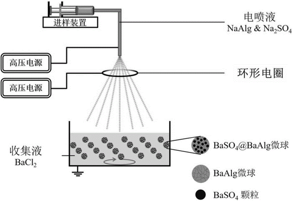
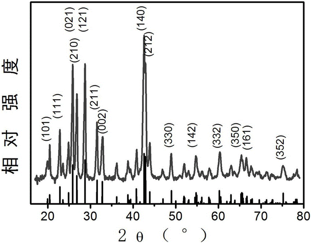
[0039] Example 1: Preparation of barium alginate microspheres loaded with barium sulfate

[0040] 2% (w / v) sodium alginate and 0.3 mol / L sodium sulfate were used as the electrospray liquid and put into the sampling device of the electrostatic spraying equipment, and the sampling speed was adjusted to 0.3 mL / hr. Use 0.6mol / L barium chloride solution as the collection liquid, place it 9cm directly below the nozzle, and slowly stir the collection liquid. The ring electric coil is placed 2cm below the nozzle to limit the spraying range of the droplets. The inner diameter of the nozzle is 0.18mm. The nozzle is connected to a DC high-voltage power supply, and the adjusted voltage is 10kv. The ring electric coil is connected to another high-voltage power supply, and the regulating voltage is 2kv. Collection dish ground wire. Turn on the DC high-voltage power supply, and the electrospray liquid is split into micron-sized droplets with uniform particle size by electrostatic force, ...

Embodiment 2

[0042] Embodiment 2: the control of developing microsphere particle size

[0043] The preparation method is consistent with Example 1. This example is only used to list some examples, showing that monodisperse microspheres with different particle sizes can be obtained by simply adjusting the parameters of electrostatic spraying, which can be applied to blood vessel embolization with different calibers.

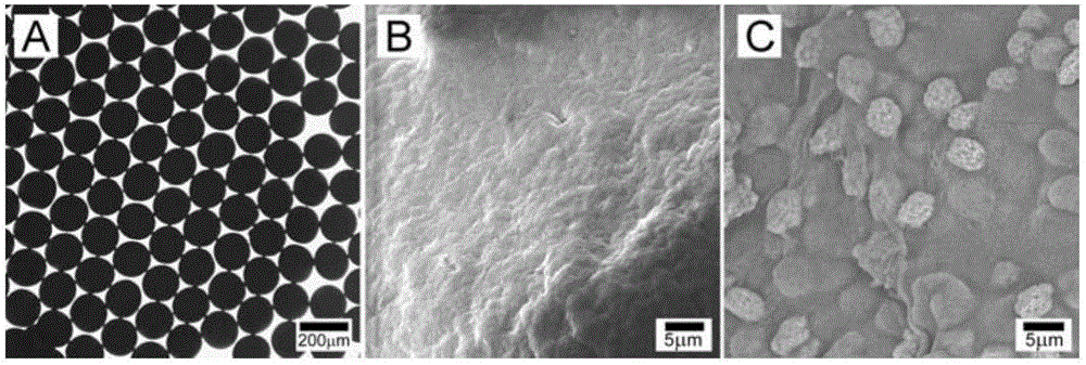
[0044] When keeping other parameters of electrostatic spraying constant and only increasing the inner diameter of the nozzle, the particle size of embolism microspheres increases accordingly. Such as Figure 4 As shown, when the inner diameter of the nozzle increases from 0.18mm, 0.26mm, 0.41mm, 0.84mm to 1.19mm, the particle size of the microspheres increases from 160±7μm, 220±18μm, 320±17μm, 410±27μm to 490 ± 23 μm. The morphology of the obtained developed microspheres is as follows: Figure 5 As shown, 5A-5E represent the optical micrographs of the developed microsphere...

Embodiment 3

[0046] Embodiment 3: Expansion of the preparation method of developing microspheres

[0047] The preparation method is consistent with Example 1. This example is only used to enumerate some examples to prove that the preparation method of the present invention is easy to expand.

[0048] Such as Figure 8 As shown, when the other parameters of electrostatic spraying are kept constant and the injection speed is increased from 0.3mL / hr, 0.6mL / hr, 1mL / hr to 2mL / hr, the particle diameters of the obtained microspheres are 160±7μm, 164 ±9 μm, 170±11 μm, and 168±9 μm. The morphology of the obtained developed microspheres is as follows: Figure 9 As shown, 9A-9D represent the optical micrographs of the microspheres prepared when the injection speed was 0.3, 0.6, 1 and 2 mL / hr, respectively. The particle size and monodispersity of the microspheres did not change much. That is, when the yield is increased by about 10 times, the quality of the developed microspheres is still within ...


PUM

PropertyMeasurementUnit
particle diameteraaaaaaaaaa
particle sizeaaaaaaaaaa
particle diameteraaaaaaaaaa

Abstract

The invention discloses a developing embolic material and a preparation method of the developing embolic material. Developable barium-alginate embolic microspheres encapsulating in-situ synthesis barium sulfate particles are prepared from one step by the adoption of an electrostatic spraying technology, integration of a developing agent and the embolic material is achieved, and the problems that when interventional therapy is clinically adopted, indirect developing exists and rechecking is difficult are solved. The mono-dispersion microspheres with the grain diameter being 100-1000 microns can be obtained by adjusting and controlling the electrostatic spraying parameter, so that the mono-dispersion microspheres with the grain diameter being 100-1000 microns are suitable for embolism of vessels of different calibers. The development agent, barium sulfate, and the barium-alginate microspheres are formed at the same time, and the barium sulfate particles are evenly distributed in the microspheres and firmly fixed. Extracorporeal simulation experiments prove that the developing microspheres are quite stable within the testing time of 50 days. A big-eared rabbit right kidney arterial embolism experiment proves that the developing microspheres have a good developing effect and an embolic effect. Because the electrostatic spraying technology has the characteristics of being simple, rapid and low in cost, the one-step preparation method has the production potential.

Description

technical field [0001] The invention relates to a preparation method of developable embolic microspheres, in particular to one-step preparation of imageable barium alginate embolic microspheres carrying in-situ synthesized barium sulfate particles by electrostatic spraying technology. Background technique [0002] Vascular interventional therapy is carried out under the guidance and monitoring of imaging equipment throughout the whole process. With the help of catheters, needles and other instruments inserted into the viscera, embolic materials are injected into the blood vessels of diseased organs to interrupt blood flow, block the supply of nutrients to the diseased part, and inhibit tumors. grow. It has the advantages of accurate and direct access to the lesion, small wound, low risk, and quick recovery, and has been widely used in tumor embolization therapy. [0003] However, the technology has the following problems in clinical use: [0004] (1) Lack of autographic em...

Claims


Application Information

Patent Timeline
Patent Type & Authority Applications(China)
IPC IPC(8): A61L24/08A61L24/02
CPCA61L24/001A61L24/0031A61L24/02A61L24/08C08L5/04
Inventor 杨祥良杜青李玲郑传胜刘宏
Owner HUAZHONG UNIV OF SCI & TECH
Why Eureka
Try Eureka