
Sequence slice-based microscope image acquisition method

A technology of image acquisition and microscopy, which is applied in the fields of instruments, character and pattern recognition, computer components, etc.

Active Publication Date: 2017-04-19
INST OF AUTOMATION CHINESE ACAD OF SCI
View PDF7 Cites 9 Cited by
  • Summary
  • Abstract
  • Description
  • Claims
  • Application Information

AI Technical Summary

Problems solved by technology

[0005] The embodiment of the present invention provides a microscope image acquisition method based on serial slices to solve the technical problem of how to efficiently complete the automatic acquisition of images of sample regions of interest

Method used


Image

  • Sequence slice-based microscope image acquisition method
  • Sequence slice-based microscope image acquisition method
  • Sequence slice-based microscope image acquisition method

Examples


Embodiment Construction

[0064] The present invention will be described in detail below in conjunction with the accompanying drawings and specific embodiments.

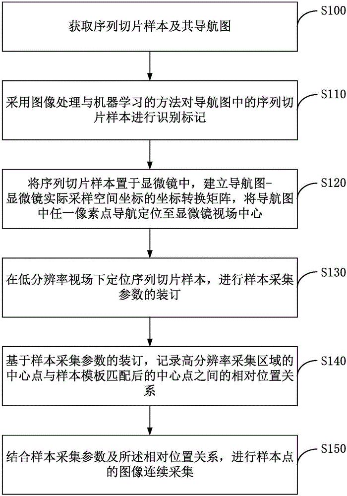
[0065] Aiming at the automatic acquisition of microscope images of serial slices, the embodiment of the present invention proposes a microscope image acquisition method based on serial slices, such as figure 1 As shown, the method may include: step S100 to step S150. in:

[0066] S100: Obtain a sequence slice sample and its navigation map.

[0067] Among them, the acquisition of the navigation map can be obtained by taking a complete photo with an optical camera, or taking partial images through a high-resolution microscope, and then splicing them into a complete navigation map.

[0068] S110: Using image processing and machine learning methods to identify and mark sequence slice samples in the navigation map.

[0069] This step records the coordinate point position of each sequence slice sample in the navigation map, and then can determin...


PUM


Abstract

The present invention discloses a sequence slice-based microscope image acquisition method. The method includes the following steps that: sequence slice samples and the navigation chart thereof are acquired; an image processing and machine learning method is adopted to identify and mark the sequence slice samples in the navigation chart; the sequence slice samples are arranged in a microscope, any pixel in the navigation chart is navigated to the center of the field of view of the microscope; the sequence slice samples are positioned under a low-resolution field of view, and binding is performed on sample acquisition parameters; and based on the binding of the sample acquisition parameters, the relative position relation of the center point of a high-resolution acquisition region and the matched center point of a sample template is recorded; and sample point image continuous image acquisition is performed based on the sample acquisition parameters and the relative position relation. With the sequence slice-based microscope image acquisition method of the technical schemes of the invention adopted, the automatic acquisition of the images of sample regions of interest can be completed efficiently, and the automatic imaging of the continuous regions of large-scale sequence samples under a condition that a microscope (an electronic microscope or optical microscope) with an automatic control interface is adopted can be realized.

Description

technical field [0001] Embodiments of the present invention relate to the field of imaging technology, and in particular to a method for acquiring microscope images based on sequential slices. Background technique [0002] Ultra-thin serial section 3D imaging technology refers to the technology of cutting biological tissue into ultra-thin serial sections or slices, and using electron microscope to image them one by one; according to different segmentation techniques, it is mainly divided into serial section imaging and serial slice imaging. Types: (1) Sequential cross-sectional imaging is the combination of slicing and imaging in the sample cavity of the electron microscope. This type of method has close resolution in the horizontal and vertical directions and is isotropic. The disadvantage lies in the destructive slicing method. Once the data is found (2) Sequential slice imaging method cuts the sample into serial slices using an ultra-thin section device, collects them wit...

Claims


Application Information

Patent Timeline
IPC IPC(8): G06K9/00G06K9/46G06K9/62
CPCG06V20/695G06V10/443G06F18/23213G06F18/214
Inventor 李国庆马宏图韩华魏利新
Owner INST OF AUTOMATION CHINESE ACAD OF SCI
Learn More