
A system and method for detecting the definition of digital pathological slides

A digital pathology slice and digital pathology technology, applied in the field of medical image detection, can solve the problems of time-consuming, difficult to measure the standard of manual image evaluation, and large digital slices, so as to reduce workload and avoid inconsistent evaluation results

Active Publication Date: 2021-05-11
KONFOONG BIOTECH INT
View PDF8 Cites 0 Cited by
  • Summary
  • Abstract
  • Description
  • Claims
  • Application Information

AI Technical Summary

Problems solved by technology

But the digitized slice is too large, so it will take more time to evaluate the whole slice
Moreover, the standard of manual image evaluation is difficult to measure, and it is strongly influenced by each person's subjective factors. The results of the same slice will be different for each person.

Method used


Image

  • A system and method for detecting the definition of digital pathological slides
  • A system and method for detecting the definition of digital pathological slides
  • A system and method for detecting the definition of digital pathological slides

Examples


Embodiment Construction

[0037] The present invention will be further described below in conjunction with the accompanying drawings and specific embodiments, but not as a limitation of the present invention.

[0038] Digital pathological slice is to scan the traditional glass pathological slice through automatic microscope or optical magnification system to obtain high-resolution digital images, and then apply computer to automatically stitch and process the obtained images with high precision and multi-view seamlessly to produce a whole full view digitized slices. Therefore, digital pathology slides are seamlessly spliced ​​and processed from multiple digital pathology images.

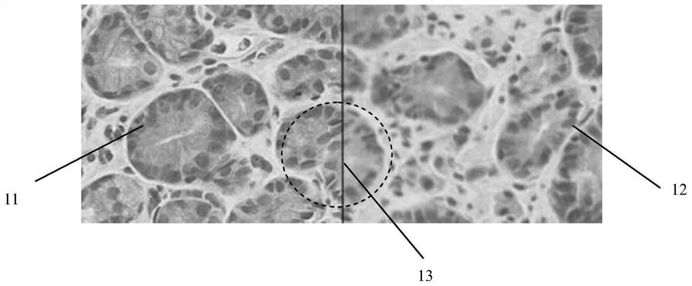
[0039] Listed in the present invention by figure 1 A digital pathological slice spliced ​​from one digital pathological picture 11 and another digital pathological picture 12 is shown as an example to illustrate the solution of the present invention.

[0040] The one digital pathological picture 11 and another digital patho...


PUM


Abstract

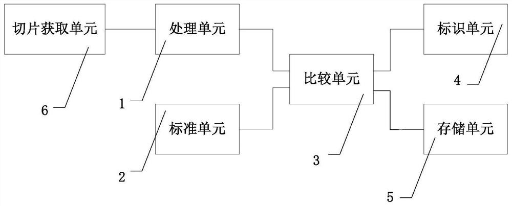
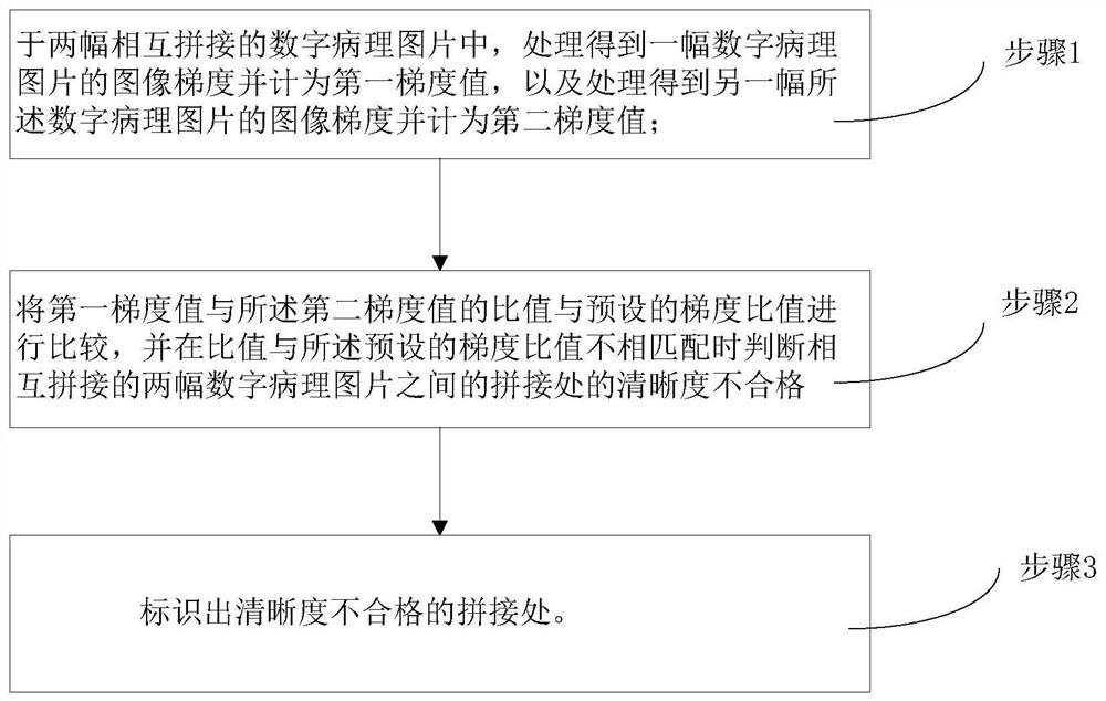
The invention relates to the field of medical image detection, in particular to a digital pathological slice definition detection system and method. The digital pathological slice includes at least a plurality of digital pathological pictures that are spliced ​​with each other, and the detection system includes: a processing unit that processes the two digital pathological pictures that are spliced ​​together to obtain an image gradient of a digital pathological picture and counts it as the first gradient value, And process the image gradient of another piece of digital pathology picture and count it as the second gradient value; the standard unit provides a preset gradient ratio in advance; the comparison unit compares the ratio of the first gradient value to the second gradient value with the preset Compare the gradient ratios of the two digital pathology pictures, and judge that the clarity of the splicing between the two digital pathology pictures that are spliced ​​with each other is unqualified when the ratio does not match the preset gradient ratio. This method can objectively judge the image quality of the image, and not only avoids the disadvantage of inconsistent evaluation results caused by human subjective factors.

Description

technical field [0001] The invention relates to the field of medical image detection, in particular to a system and method for detecting the definition of digital pathological slices. Background technique [0002] Digital pathological slides are a technology that combines modern digital systems with traditional optical magnification devices. It scans the traditional glass pathological sections through a fully automatic microscope or an optical magnification system to obtain high-resolution digital images, and then uses a computer to automatically stitch and process the obtained images with high precision and multi-view seamlessly to produce a digital image of the entire full view. slice. [0003] Because the high-resolution digital images scanned by the microscope or the optical magnification system are easily affected by the surrounding environment or the microscope's own equipment, the resolution of each scanned digital image is different, and some digital images are very...

Claims


Application Information

Patent Timeline
Patent Type & Authority Patents(China)
IPC IPC(8): G06T7/00
CPCG06T7/0014G06T2207/10056G06T2207/30004
Inventor 刘炳宪谢菊元王焱辉王克惠郝美蓉
Owner KONFOONG BIOTECH INT
Learn More