
An Autofocus Method Based on Image Processing in Dual-Channel Fluorescence Optical Microscopy

An optical microscope and auto-focus technology, applied in optics, microscopes, optical components, etc., can solve problems such as changes and achieve the effect of increasing stability and accuracy

Active Publication Date: 2018-10-16
HUAZHONG UNIV OF SCI & TECH
View PDF4 Cites 0 Cited by
  • Summary
  • Abstract
  • Description
  • Claims
  • Application Information

AI Technical Summary

Problems solved by technology

[0004] Aiming at the defects of the prior art, the purpose of the present invention is to provide an autofocus method based on image processing in dual-channel fluorescence optical microscopic imaging, aiming to solve the problem of imaging biological tissue samples using a fluorescent optical microscopic imaging system. The focal plane will change with the ambient temperature and the solution concentration between the objective lens and the surface of the biological tissue sample.

Method used


Image

  • An Autofocus Method Based on Image Processing in Dual-Channel Fluorescence Optical Microscopy
  • An Autofocus Method Based on Image Processing in Dual-Channel Fluorescence Optical Microscopy
  • An Autofocus Method Based on Image Processing in Dual-Channel Fluorescence Optical Microscopy

Examples


Embodiment Construction

[0031] In order to make the object, technical solution and advantages of the present invention clearer, the present invention will be further described in detail below in conjunction with the accompanying drawings and embodiments. It should be understood that the specific embodiments described here are only used to explain the present invention, not to limit the present invention.

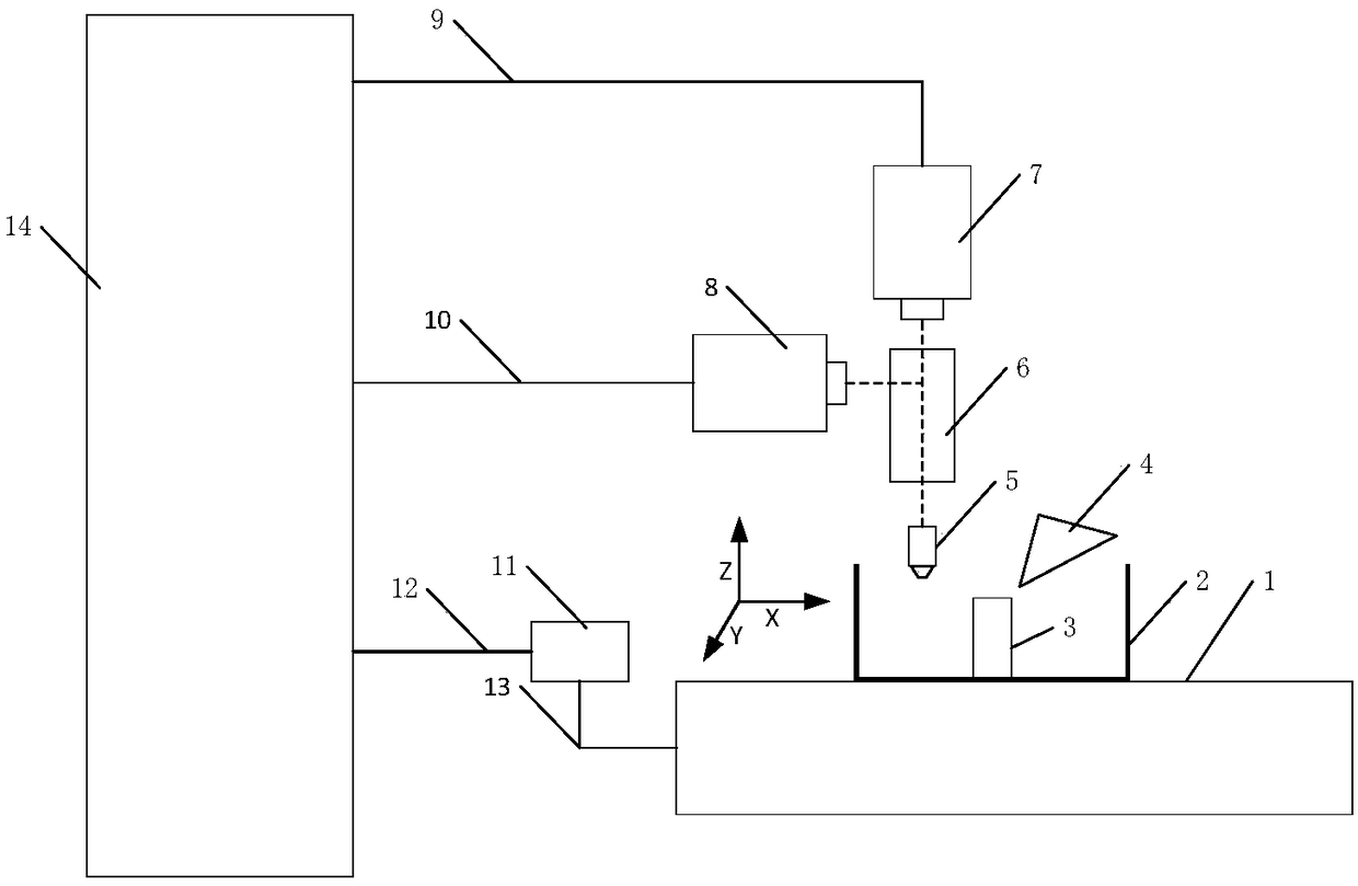
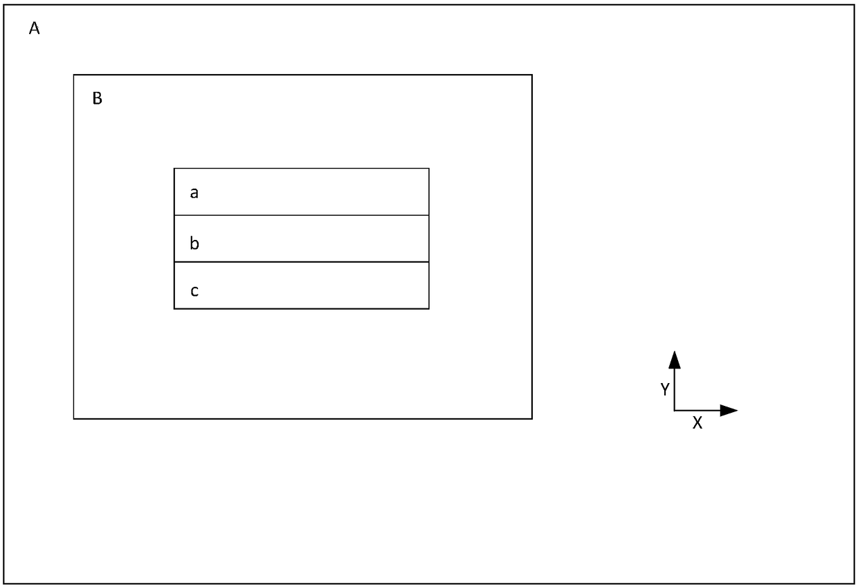
[0032] The image processing-based autofocus method in the dual-channel fluorescence microscopic imaging system provided by the present invention is suitable for systems supporting dual-channel fluorescent optical microscopic imaging; it has high focusing quality and focusing stability. In the dual-channel fluorescence imaging of biological tissue samples, one channel performs fluorescence imaging of biomarker signals, and the other channel images the cellular structure of biological tissue samples. The cell structure channel of the biological tissue sample can observe the fluorescent image of the s...


PUM


Abstract

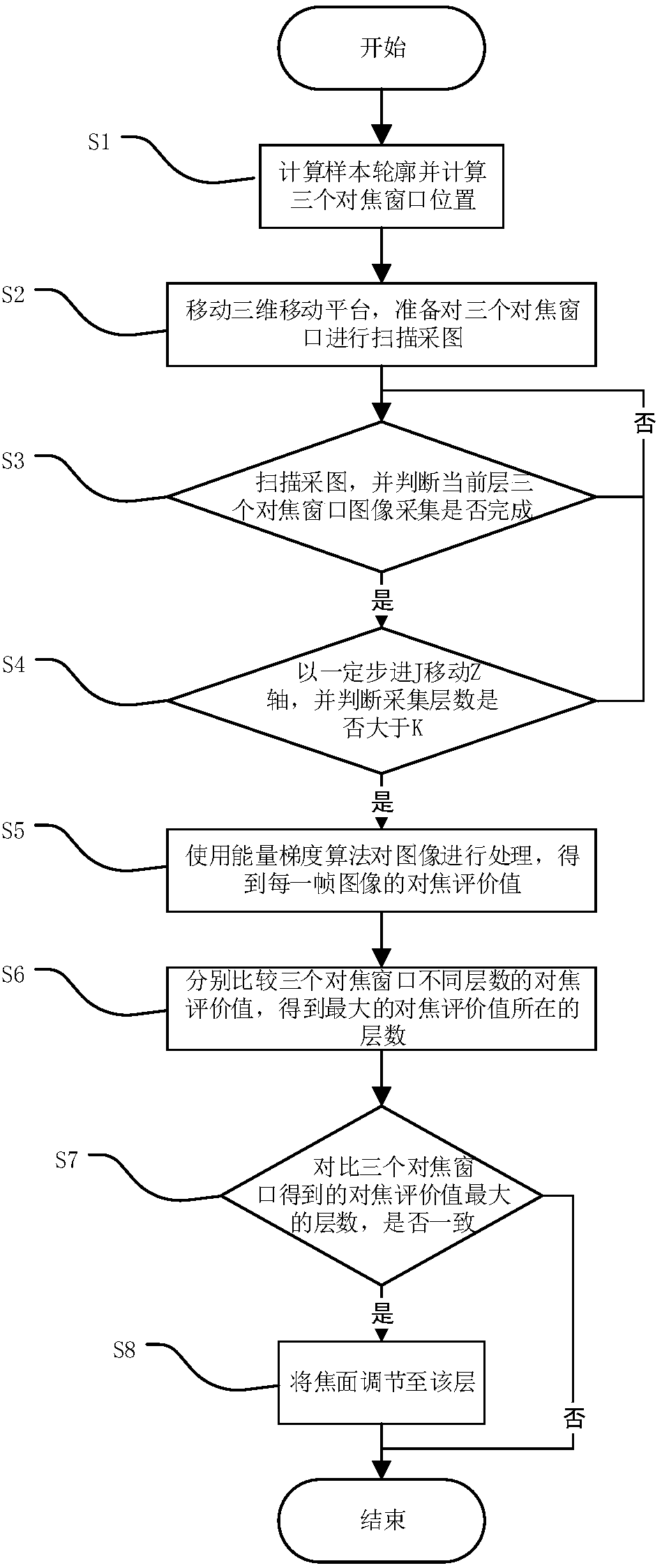
The invention discloses an image processing-based automatic focusing method applied to dual-channel fluorescence optical microscopic imaging. The method includes the following steps that: S1, the current coronal plane profile of a biological tissue sample is acquired, and three focusing window positions are obtained; S2 the three focusing windows are scanned, the image of the i-th layer of the cell construction channel of the biological tissue sample is acquired; S3 whether the image acquisition of the three focusing windows at the current layer is completed judged, if the image acquisition is completed, 1 is added to i, and the method enters S4, otherwise, the method returns to S2; S4, whether the number i of acquired layers is larger than a preset threshold value is judged, if the number i of the acquired layers is larger than the preset threshold value, the method enters S5, otherwise, the method returns to S2; S5, the acquired images of the cell construction channels of the biological tissue sample are processed, so that the focusing evaluation value of each frame of image is obtained; S6, layers of which the focusing evaluation values are maximum are obtained; S7, whether layers of which the focusing evaluation values are maximum are the same layer is judged, if the layers of which the focusing evaluation values are maximum are the same layer, the method enters S8, otherwise, the method terminates; and S8, a size to which a focal plane needs to be adjusted is obtained, and focusing is carried out according to the size to which the focal plane needs to be adjusted.

Description

technical field [0001] The invention belongs to the technical field of biological imaging, and more specifically relates to an automatic focusing method based on image processing in dual-channel fluorescence optical microscopic imaging. Background technique [0002] In fluorescence optical microscopy imaging, in order to obtain complete morphological data of biological tissue samples, the entire biological tissue sample is usually sliced ​​and imaged, and the process takes several days. Bioluminescent signals are basically extremely weak and delicate, especially nerve signals, whose size is on the order of hundreds of nanometers. In fluorescence optical microscopy imaging, there is a certain medium between the objective lens and the surface of the biological tissue sample, that is, air, buffered aqueous solution, etc. If the medium changes during the imaging process, such as the temperature and concentration of the buffered aqueous solution, it will affect the light in the w...

Claims


Application Information

Patent Timeline
Patent Type & Authority Patents(China)
IPC IPC(8): G02B21/24
CPCG02B21/244
Inventor 曾绍群吕晓华白柯尹芳芳黄凯李宁
Owner HUAZHONG UNIV OF SCI & TECH
Learn More